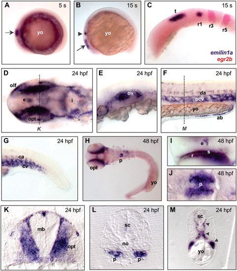
Whole-mount in situ hybridization of zebrafish embryos, visualizing the expression pattern of emilin1a at different developmental stages. A: Expression in the midbrain (arrow) at 5 somites (s). B: Expression in the midbrain (arrow) and in the first rhombomere (arrowhead) of hindbrain at 15 s. C: Double staining with egr2b probe (in red), labeling the third and fifth rhombomeres of hindbrain, confirms emilin1a expression in the dorsal side of first rhombomere and in the midbrain tectum at 15 s. D: Expression in epiphysis and head mesenchyme, especially in the territories of the olfactory and optic primordia, at 24 hours postfertilization (hpf). E: Expression in the otic vesicle at 24 hpf. F,G: Expression in the circulatory system of the trunk (F) and tail (G) at 24 hpf. H-J: Low (H) and high (I,J) magnifications of 48 hpf embryos, showing expression of emilin1a in epiphysis, sclera and retina (H,I), and in the pronephros (H,J). K: Cross-section of a 24 hpf embryo (level of section is marked by a dotted line in D), showing expression of emilin1a in the head mesenchyme. L: Cross-section of a 24 hpf embryo, showing expression in the pronephros. M: Cross-section of a 24 hpf embryo (level of section is marked by a dotted line in F), showing expression in the arterial, venous, and intersegmentary blood vessels (arrowheads). Except for embryos in panels A, B, and M, yolk was removed and embryos were mounted between glass coverslips. In A-J, anterior side of embryo is on the left; in the cross-sections, dorsal side is at the top. A-C and E-G are lateral views; D, H, and I are dorsal views; J is a ventral view. ab, angioblasts; ca, caudal aorta; cv, cardinal vein; da, dorsal aorta; e, epiphysis; i, isthmus; mb, midbrain; no, notochord; olf, olfactory placode; opt, optic cup; ov, otic vesicle; p, pronephros; pcv, posterior cardinal vein; r1-r5, first-fifth rhombomeres; r, retina; s, sclera; sc, spinal cord; t, tectum; yo, yolk.
|
