
|
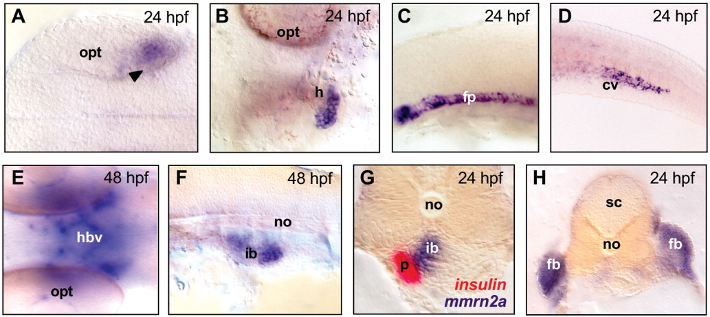
Fig. 6 Whole-mount in situ hybridization for mmrn2a in zebrafish embryos at 24 hours postfertilization (hpf) and 48 hpf. A: Expression in the posterior retina (arrowheads) of the developing eye at 24 hpf. B: Ventral view, showing expression in the heart at 24 hpf. C,D: Expression in the anterior part of the floor plate (C) and in the caudal vein (D) at 24 hpf. E: Expression in the circulatory system of the head at 48 hpf. F: Expression in a territory corresponding to the developing intestinal bulb. G: Cross-section of a 24 hpf embryo after double labeling with insulin and mmrn2a probes; the insulin probe (in red) marks the β-cell precursors of pancreas, located behind the intestinal bulb, while mmrn2a labels the intestinal bulb but not the pancreas. H: Cross-section of a 24 hpf embryo, showing expression in the fin buds. All embryos were dissected away from the yolk and flattened. In A-F, anterior side of embryo is on the left; in the cross-sections, dorsal side is at the top. A and E are dorsal views, B is a ventral view, C,D,F are lateral views. cv, caudal vein; fb, fin bud; fp, floor plate; h, heart; hbv, head blood vessels; ib, intestinal bulb; no, notochord; opt, optic cup; p, pancreas; sc, spinal cord.