|
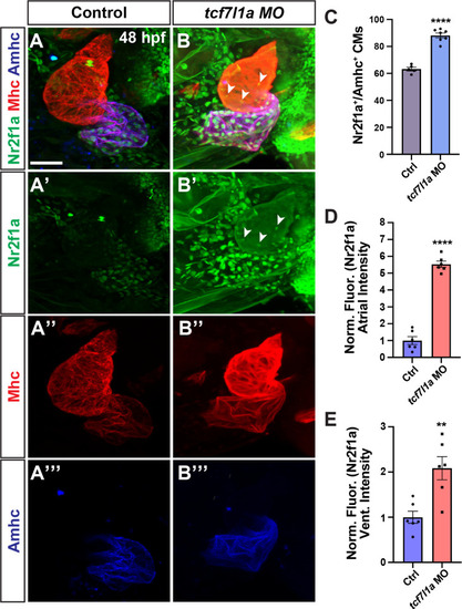
Tcf7l1a limits Nr2f1a+ cardiomyocytes within the heart. A-B”’) Confocal images of hearts from control and tcf7l1a MO-injected embryos. Nr2f1a (green), Mhc (red), Ahmc (blue). Images are frontal views with the arterial pole up. Arrowheads in B and B’ indicate Nr2f1a+ nuclei in the ventricle. Scale bar: 50 μm. C) The number of Nr2f1a+/Amhc+ cardiomyocytes in the hearts of control and tcf7l1a MO-injected embryos. Control (n = 6); tcf7l1a MO-injected (n = 7) embryos. D) Normalized intensity of Nr2f1a expression in atria of control and tcf7l1a MO-injected embryos. Control (n = 6); tcf7l1a MO-injected (n = 6) embryos. E) Normalized intensity of Nr2f1a expression in ventricles of control and tcf7l1a MO-injected embryos. Control (n = 6); tcf7l1a MO-injected (n = 6) embryos. ** indicates P = 0.0038; **** indicates P< 0.0001.
|