|
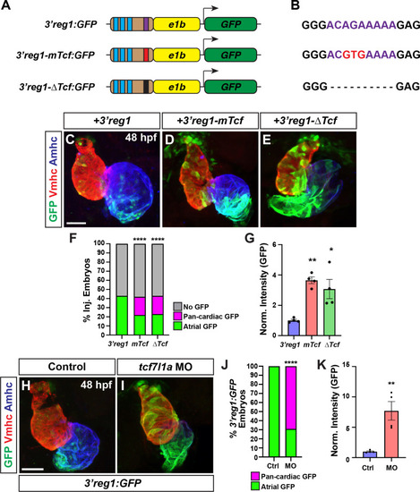
Tcf7l1a restricts 3’reg1 reporter expression within the heart. A) Schematics of 3’reg1:GFP reporter constructs. Foxf sites (blue), Lef/Tcf site (purple), mutated Lef/Tcf site (red), and deleted Lef/Tcf site (black). B) Sequences of the WT (purple), mutated (red), and deleted Left/Tcf sites within the 3’reg1 enhancer. C-E) Confocal images of hearts from embryos injected with the 3’reg1:GFP, 3’reg1-mTcf:GFP, 3’reg1-ΔTcf:GFP constructs. F) The percentage of transient transgenic embryos with reporter atrial, pan-cardiac, and lacking expression in their hearts. 3’reg1:GFP (n = 200); 3’reg1-mTcf:GFP (n = 101); 3’reg1-ΔTcf:GFP (n = 102). G) Normalized intensity of GFP expression in hearts from 3’reg1:GFP (n = 4), 3’reg1-mTcf:GFP (n = 4), and 3’reg1-ΔTcf:GFP (n = 4) embryos. H,I) Confocal images of hearts from control and tcf7l1a MO-injected transgenic 3’reg1:GFP embryos. J) The percentage of stable 3’reg1:GFP embryos with reporter atrial and pan-cardiac expression in their hearts. Control (n = 152); tcf7l1a-MO (n = 92). Images are frontal views with the arterial pole up. Hearts are stained for 3’reg1:GFP (green), Vmhc (red), Amhc (blue). Scale bars: 50 μm. K) Normalized intensity of GFP expression in hearts from control and tcf7l1a MO-injected stable 3’reg1:GFP embryos. Control (n = 4), tcf7l1a MO (n = 4).* indicate P < 0.02, ** indicate P<0.003, **** indicate P < 0.0001.
|