
FIGURE
Fig. 8
Fig. 8
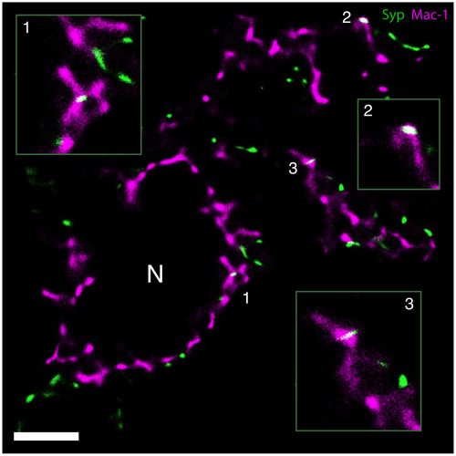
GSDIM demonstrates Syp‐immunoreactive presynaptic material (green) inside perineuronal microglial processes (Mac‐1, magenta) in the axotomized mouse facial nucleus 3 days following axotomy. Examples of stripped synapses (white, merge color) are magnified in the insets. Numbered regions represent the magnified areas in the insets. N marks a cross‐sectioned motor neuron. Scale bar = 2.5 µm |
Expression Data
Expression Detail
Antibody Labeling
Phenotype Data
Phenotype Detail
Acknowledgments
This image is the copyrighted work of the attributed author or publisher, and
ZFIN has permission only to display this image to its users.
Additional permissions should be obtained from the applicable author or publisher of the image.
Full text @ J. Neurosci. Res.