
IMAGE
Fig. 8
- ID
- ZDB-IMAGE-210518-8
- Publication
- Paasila et al., 2021 - Ground state depletion microscopy as a tool for studying microglia-synapse interactions
- All Figures
- Figures for Paasila et al., 2021
Image
|
Figure Caption
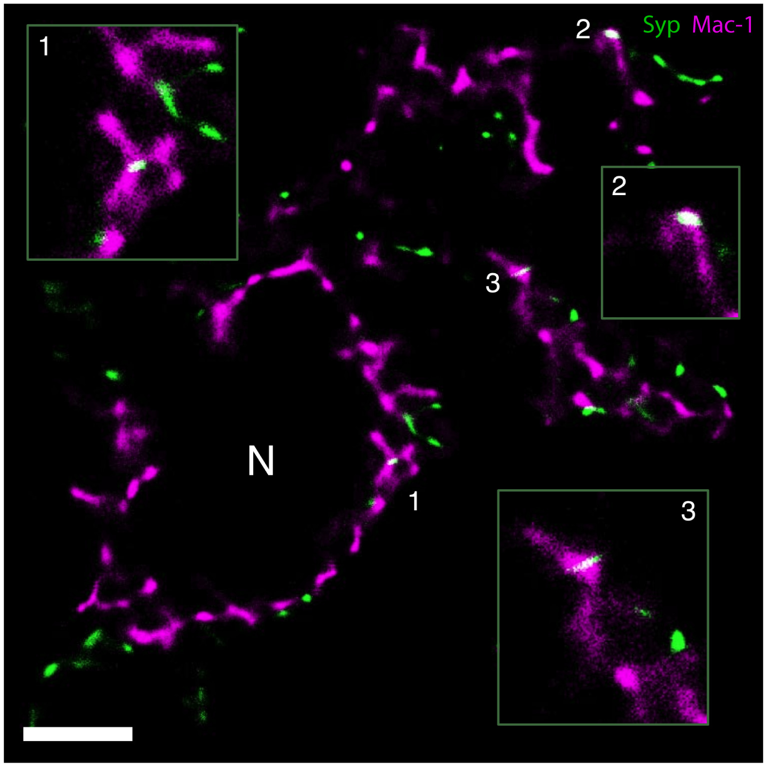
Fig. 8 GSDIM demonstrates Syp‐immunoreactive presynaptic material (green) inside perineuronal microglial processes (Mac‐1, magenta) in the axotomized mouse facial nucleus 3 days following axotomy. Examples of stripped synapses (white, merge color) are magnified in the insets. Numbered regions represent the magnified areas in the insets. N marks a cross‐sectioned motor neuron. Scale bar = 2.5 µm
Acknowledgments
This image is the copyrighted work of the attributed author or publisher, and
ZFIN has permission only to display this image to its users.
Additional permissions should be obtained from the applicable author or publisher of the image.
Full text @ J. Neurosci. Res.