|
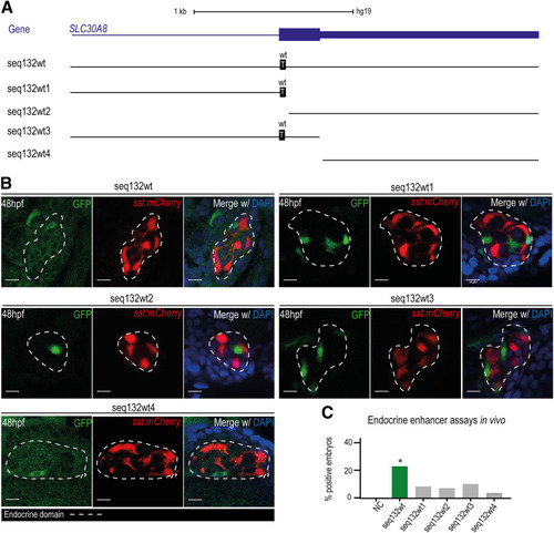
A: Schematic representation of the four analyzed fragments derived from seq132wt: seq132wt1, seq132wt2, seq132wt3, and seq132wt4. The gene SLC30A08 is represented in blue. The wt allele is discriminated in black boxes. B: Representative confocal images for the total sequence, seq132wt, and the four analyzed fragments showing GFP-positive cells in the endocrine pancreatic domain (dashed line) defined by the sst:mCherry reporter line. The 48-hpf embryos were stained with DAPI. Scale bars = 10 µm. C: Graph representing the total percentages of positive embryos for seq132wt (23%, n = 34), seq132wt1 (9.8%, n = 41), seq132wt2 (6.5%, n = 31), seq132wt3 (10%, n = 30), and seq132wt4 (4%, n = 23). *P < 0.05, by χ2 test.
|