IMAGE

Figure 5

ID
ZDB-IMAGE-201208-37
Source
Figures for Eufrásio et al., 2020
Image
Figure Caption

Figure 5

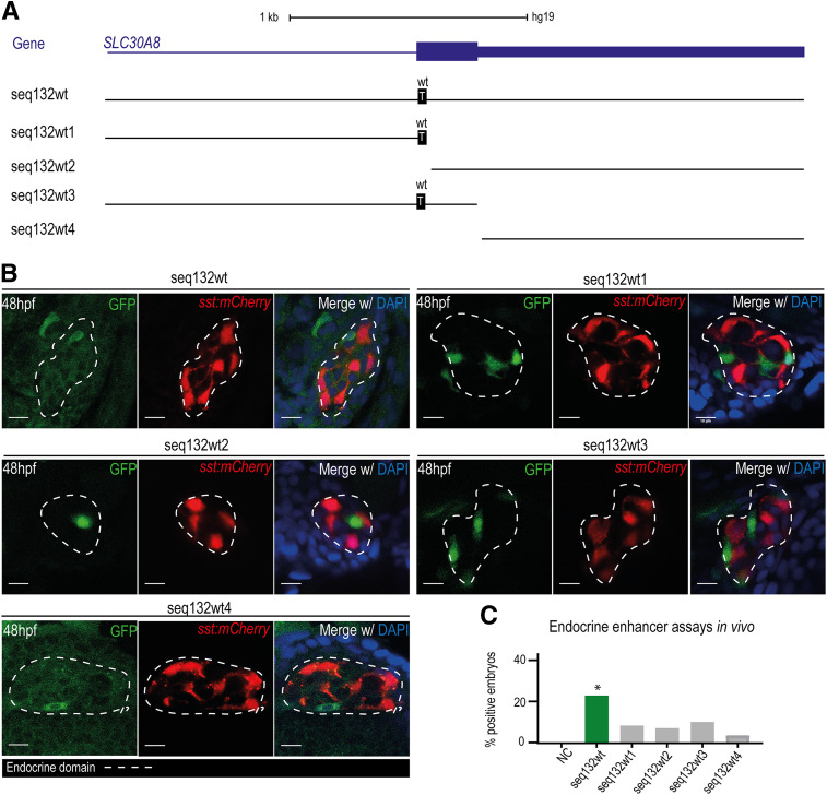
A: Schematic representation of the four analyzed fragments derived from seq132wt: seq132wt1, seq132wt2, seq132wt3, and seq132wt4. The gene SLC30A08 is represented in blue. The wt allele is discriminated in black boxes. B: Representative confocal images for the total sequence, seq132wt, and the four analyzed fragments showing GFP-positive cells in the endocrine pancreatic domain (dashed line) defined by the sst:mCherry reporter line. The 48-hpf embryos were stained with DAPI. Scale bars = 10 µm. C: Graph representing the total percentages of positive embryos for seq132wt (23%, n = 34), seq132wt1 (9.8%, n = 41), seq132wt2 (6.5%, n = 31), seq132wt3 (10%, n = 30), and seq132wt4 (4%, n = 23). *P < 0.05, by χ2 test.

Acknowledgments
This image is the copyrighted work of the attributed author or publisher, and ZFIN has permission only to display this image to its users. Additional permissions should be obtained from the applicable author or publisher of the image. Full text @ Diabetes