FIGURE

Figure 2

ID
ZDB-FIG-200212-34
Publication
Ki et al., 2019 - CEP41-mediated ciliary tubulin glutamylation drives angiogenesis through AURKA-dependent deciliation
Other Figures
All Figure Page
Back to All Figure Page
Figure 2

Ablation of <italic>cep41</italic> impairs vascular development in zebrafish

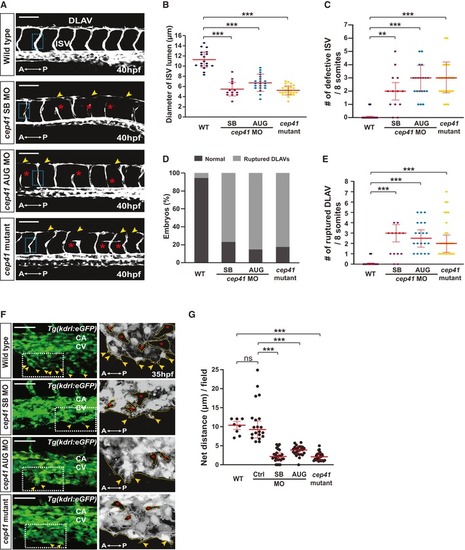
Blood vessels were observed in MOs (control, cep41 AUG (2.5 ng), or cep41 SB (2 ng))‐injected or cep41‐mutated Tg(kdrl:eGFP) zebrafish at 40 hours post‐fertilization (hpf) by fluorescent microscopy. Asterisks and arrowheads indicate impaired ISVs and DLAVs, respectively. The representative images for analysis of ISV lumen diameter are indicated by dotted rectangles. A, anterior; P, posterior; DLAV, dorsal longitudinal anastomotic vessel; ISV, intersegmental vessel. Scale bars, 100 μm.

Quantification of ISV lumen diameter in (B), the numbers of defective ISVs in (C), the numbers of embryos with aberrant DLAVs in (D), and the numbers of ruptured DLAVs in (E) from data observed in equivalent fields of view (within eight somites). The severity of blood vessel defects in cep41‐deficient zebrafish: B (narrowed ISVs) < C (shorten, fused, and missing ISVs) < D and E (ruptured DLAVs). Data are shown as mean ± SD of three independent experiments with ≥ 20 embryos per condition. Statistical significance was determined using the one‐way ANOVA followed by Dunnett's post hoc test (B) and the Kruskal–Wallis test by Dunn's post hoc test (C, E) (**< 0.01, ***< 0.001).

CV protrusions (arrowheads) and vascular loops (asterisks) were observed in zebrafish at 35 hpf. The images within the dotted rectangles are magnified in the right panels. A, anterior; P, posterior; CA, caudal artery; CV, caudal vein. Scale bars, 40 μm. Quantification of the length of the CV protrusions in (G) presented graphically by measuring them in equivalent fields of view (within five somites). Data are median of four independent experiments ( 5 embryos per experimental condition). Statistical significance was determined using the Kruskal–Wallis test followed by Dunn's post hoc test (***< 0.001, ns: non‐significant).

Expression Data

Expression Detail
Antibody Labeling
Phenotype Data
Fish:
Knockdown Reagents:
Observed In:
Stage Range: Prim-15 to Prim-25

Phenotype Detail
Acknowledgments
This image is the copyrighted work of the attributed author or publisher, and ZFIN has permission only to display this image to its users. Additional permissions should be obtained from the applicable author or publisher of the image. Full text @ EMBO Rep.