<styled-content toggle='no' style='fixed-case'>CEP</styled-content>41 facilitates tubulin glutamylation in endothelial cilia
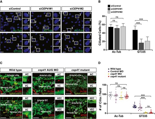
Control or CEP41 siRNA‐transfected HUVECs were immunostained with Ac‐Tub‐ or GT335‐specific antibodies in (A). The rectangles indicate the representative cells from each immunostaining experiment presented as magnified images in the right panels. Scale bars, 20 μm. Quantification of the ciliated cell numbers in the images in (A) from the results of three independent experiments with ≥ 200 cells per condition (mean ± SD) in (B). ***P <0.001, ns: non‐significant (one‐way ANOVA with Dunnett's post hoc test).
Wild‐type and cep41‐deficient Tg(kdrl:eGFP) zebrafish embryos were fixed for immunostaining with Ac‐Tub‐ or GT335‐specific antibodies at 28 hpf in (C). The rectangles indicate the arterial cilia (AC) and venous cilia (VC) in zebrafish endothelial cells (EC). Magnified representative images are displayed in the right panels. Scale bars, 40 μm. Quantification of the labeled cilia observed in equivalent fields of view is presented graphically in (D). Data are shown as median of three independent experiments (n ≥20 embryos per condition). Statistical significance was determined using the Kruskal–Wallis test followed by Dunn's post hoc test (***P <0.001, ns: non‐significant).
This image is the copyrighted work of the attributed author or publisher, and
ZFIN has permission only to display this image to its users.
Additional permissions should be obtained from the applicable author or publisher of the image.
Full text @ EMBO Rep.
Your Input Welcome
Thank you for submitting comments. Your input has been emailed to ZFIN curators who may contact you if
additional information is required.
Oops. Something went wrong. Please try again later.