
Fig. S14
- ID
- ZDB-FIG-180906-27
- Publication
- Mochizuki et al., 2017 - Cell division and cadherin-mediated adhesion regulate lens epithelial cell movement in zebrafish
- Other Figures
- All Figure Page
- Back to All Figure Page
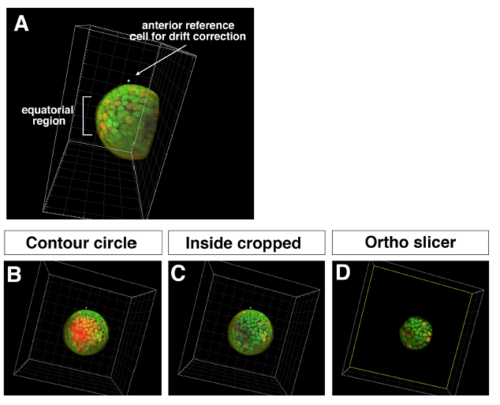
Procedure for obtaining 3D time-lapse images of equatorial lens epithelium (A) Side view of the lens scanned from the ventral view. Tissues outside the lens sphere, such as the retina and the cornea, were removed by cropping. Anterior reference position used for drift correction and the equatorial region are indicated. (B) Equatorial view of the lens before cropping the lens fiber core. Contour of the surface masking the lens fiber core is indicated (red). (C) Equatorial view of the lens after cropping the lens fiber core. (D) Only the equatorial lens epithelium was extracted using the “Ortho slicer” tool. |