
Fig. 6
- ID
- ZDB-FIG-180906-16
- Publication
- Mochizuki et al., 2017 - Cell division and cadherin-mediated adhesion regulate lens epithelial cell movement in zebrafish
- Other Figures
- All Figure Page
- Back to All Figure Page
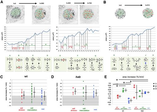
Temporal profile of lens epithelial expansion of the wild type and the E-cadherin mutant. (A) Wild-type lens epithelial area, consisting of 12 cells during the first 390 min (left panels) and 10 cells during the last 315 min (right panels). The top panels indicate the composition of individual lens epithelial cells. Divided daughter cells are in red. Middle panels display temporal profiles of the area. Red and green arrows indicate cell division events and periods when cell intercalation occurred, respectively. Bottom panels indicate cell intercalation that occurred in the corresponding period of middle panels. (B) Temporal change of habrk3 mutant lens epithelial area consisting of 14 cells during the first 315 min. (C,D) Expansion rate per time frame of lens epithelial area of wild-type (C) and habrk3 mutant (D) lens shown in A and B, respectively, during periods of cell division (red), cell intercalation (green) and the rest period (blue). Averages and s.d. are indicated by horizontal and vertical bars, respectively. The average value of the cell intercalation period is higher than others in wild type, whereas average values are similar during all three periods in habrk3 mutant. (E) The average rate of lens epithelial expansion in three wild-type and habrk3 mutant lenses. Averages and s.d. are indicated by horizontal and vertical bars, respectively. Although values of cell division period in the habrk3 mutant are variable, profiles were similar to those shown in C and D. Student's t-test: *P<0.05. |
| Fish: | |
|---|---|
| Observed In: | |
| Stage: | Long-pec |