
Fig. 3
- ID
- ZDB-FIG-170825-5
- Publication
- Paksa et al., 2016 - Repulsive cues combined with physical barriers and cell-cell adhesion determine progenitor cell positioning during organogenesis
- Other Figures
- All Figure Page
- Back to All Figure Page
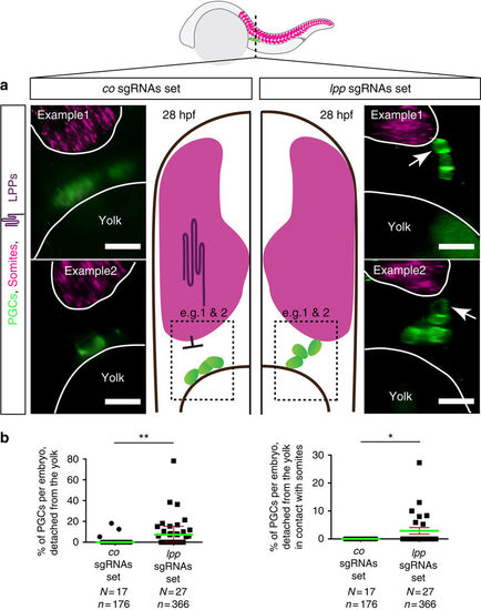
Abnormal positioning of PGCs in embryos treated with Cas9 and sgRNAs set against lpps. (a) Optical cross-sections (plane marked by dashed line in the embryo scheme) of whole-mount 28 hpf embryos (Tg(kop:egfp-f-3′nos3) expressing EGFP in their PGCs following RNAscope procedure labelling myoD expression in somites (magenta, border marked in white). In contrast with control embryos (left panels), in embryos treated with Cas9 and a set of sgRNAs targeting 6 lpps (right panels) PGCs detach from the yolk and can contact somites (arrows). Scale bars, 20 μm. Dorsal is up. (b) The percentages of PGCs per embryo detached from the yolk (left graph) and percentage of those in contact with the somites (right graph) is significantly elevated in LPPs-depleted embryos. The statistical significance was evaluated using the Mann–Whitney U-test (*P≤0.05, **P≤0.01). Green lines signify the mean, error bars the standard error of the mean (s.e.m), N the number of embryos and n the number of PGCs examined. |