
Fig. S1
- ID
- ZDB-FIG-170825-9
- Publication
- Paksa et al., 2016 - Repulsive cues combined with physical barriers and cell-cell adhesion determine progenitor cell positioning during organogenesis
- Other Figures
- All Figure Page
- Back to All Figure Page
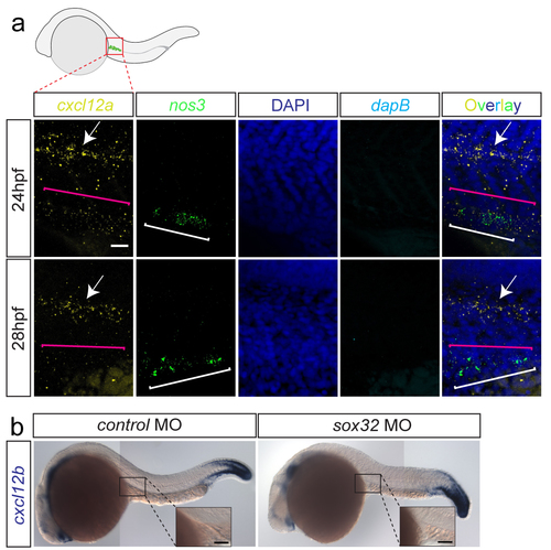
cxcl12a and cxcl12b expression patterns following PGC arrival at the site where the gonad develops (a) cxcl12a expression at the gonad region of 24hpf-embryos is detected (using the sensitive RNAscope protocol, magenta bracket in the upper panel), along with much higher expression level in the lateral line (arrow). At 28hpf, only weak background level cxcl12a is detected where the PGCs reside. PGCs detected based on nanos3 (nos3) expression (white brackets). Nuclei stained with DAPI. dapB serves as a negative control. Merged overlay images presented on the right. Scale bar 25μm. Dorsal up. (b) cxcl12b RNA expression is not detectable at the gonad region in 24hpf control embryos, nor in embryos lacking the gut tube. Insets display a higher magnification of the gonad region. Scale bars 50μm. |