
Fig. S10
- ID
- ZDB-FIG-170825-15
- Publication
- Paksa et al., 2016 - Repulsive cues combined with physical barriers and cell-cell adhesion determine progenitor cell positioning during organogenesis
- Other Figures
- All Figure Page
- Back to All Figure Page
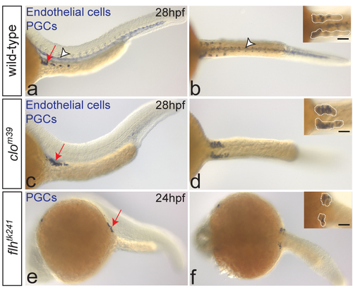
Bilateral positioning of PGC clusters does not require the notochord or blood vessles. One-color in situ hybridization on wild-type (a-b), clom39 (a-d) and flhtk241 (e-f) embryos. PGCs are stained with nos3 (a-f, arrows on the left-panel images) and endothelial cells with kdrl probe (a-d, arrowheads in a-b). Despite the lack of endothelial cells, PGCs arrive at the target (c, arrow) and form two separate clusters at 28hpf (d; dotted lines in inset) in clo mutant embryos (number of embryos examined: N=12), similar to wild-type embryos (a-b; N=60). In embryos lacking notochord (flhtk241), PGCs form two bilateral clusters at the gonad region at 24hpf (e-f; dotted lines in inset; N=15). Scale bars 50μm. Lateral (left panels) and dorsal (right panels) views are shown. |