
Fig. 1
- ID
- ZDB-FIG-170825-3
- Publication
- Paksa et al., 2016 - Repulsive cues combined with physical barriers and cell-cell adhesion determine progenitor cell positioning during organogenesis
- Other Figures
- All Figure Page
- Back to All Figure Page
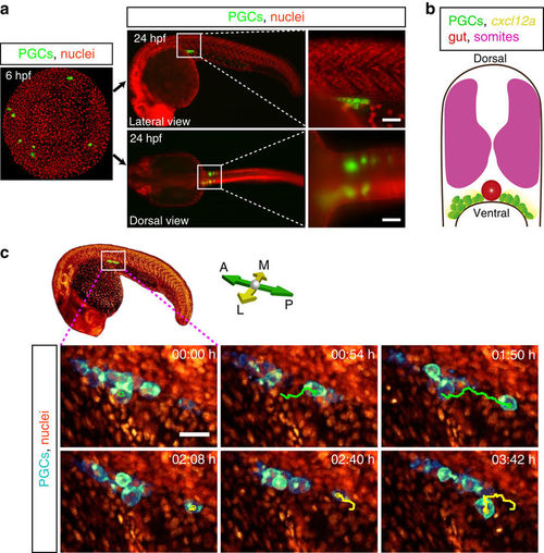
PGCs are motile at the gonad region. (a) PGCs migrate from four different positions in the embryo (green clusters in 6 hpf image) towards the developing gonads to form two separate cell clusters by end of the first day of embryonic development (lateral and dorsal views). Insets display higher magnification of the gonad region (white boxes). Scale bars represent 50 μm. (b) A schematic cross-section of a 1-day-old zebrafish embryo showing the somites (magenta), the two separate PGC clusters (green cells) located on either side of the developing gut (red structure), as well as the expression of cxcl12a at this stage (yellow). (c) Snapshots from a time-lapse movie (Supplementary Movie 2) showing a lateral view of a PGC cluster starting at 24 hpf In the first three time points posterior migration of a PGC is highlighted (green track) and lateral–medial migration of the same PGC is presented in the following panels (yellow track). Scale bar, 25 μm. |