
Fig. S12
- ID
- ZDB-FIG-170825-17
- Publication
- Paksa et al., 2016 - Repulsive cues combined with physical barriers and cell-cell adhesion determine progenitor cell positioning during organogenesis
- Other Figures
- All Figure Page
- Back to All Figure Page
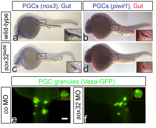
PGCs are normally specified in embryos lacking the gut tube One color (a and c) or two color (b and d) WISH on wild-type and sox32ta56 embryos at 24hpf (a and c respectively) or 28hpf (b and d respectively). The gut is stained using foxa1 probe (a-d). In wild-type and sox32 mutant embryos the PGCs (insets) express both the early marker nos3 and the late marker piwil1. Arrowheads indicate the position of the gut (a-b), and no foxa1 expression is detected in the mutants (c-d). Number of embryos examined in a: N=40, b: N=20, c: N=9 and d: N=5. (e-f) PGCs are labeled with vasa-gfp mRNA, showing the characteristic granule formation in PGCs (insets in e and f respectively). e: N=14 and f: N=15. Scale bar 50μm. Dorsal views are shown. Anterior is up. |