
Fig. S2
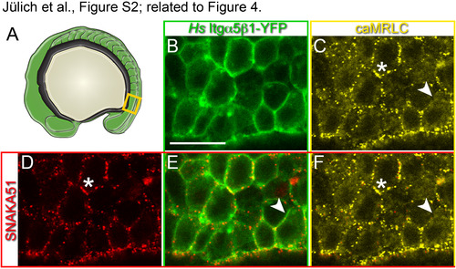
In vivo visualization of the extended, active conformation of Itgα5β1. FN matrix fibrillogenesis and Itgα5β1-FN binding is stimulated by cytoskeletal contractility (Friedland et al., 2009; Schwarzbauer and DeSimone, 2011). To validate the specificity of the SNAKA51 antibody for the activated Itgα5 conformation within the zebrafish presomitic mesoderm, we confirmed that increasing actomyosin contractility elevated SNAKA51 binding. We genetically increased actomyosin contractility by mosaically expressing an RFP-tagged activated myosin regulatory light chain 2 (caMRLC2) in MZ itgα5-/- embryos also expressing Hs Itgα5β1-YFP (Iwasaki et al., 2001; Norden et al., 2009).(A) Itgα 5 conformation was assayed in the mesenchymal presomitic mesoderm (orange box). (B) Expression of Hs Itgα5β1-YFP (green) outlines the cell cortices in the presomitic mesoderm. Scale bar is 20 µm. (C) Mosaic expression of caMRLC-RFP (yellow). (D) SNAKA51 immunolocalization (red). (E) SNAKA51 localizes with some but not all Hs Itgα5β1-YFP. (F) Puncta of caMRLC colocalize with SNAKA51 (asterisks). Arrowheads indicate inactive Itgα5β1 lacking colocalized caMRLC. In 4 experiments, 58% of 33 embryos exhibited the correlation between caMRLC expression and increased SNAKA51 signal. Thus, caMRLC2 increased SNAKA51 signal relative to adjacent control cells which is consistent with SNAKA51 recognizing the extended active form of Itgα5 in the zebrafish embryo. |
Reprinted from Developmental Cell, 34(1), Jülich, D., Cobb, G., Melo, A.M., McMillen, P., Lawton, A.K., Mochrie, S.G., Rhoades, E., Holley, S.A., Cross-Scale Integrin Regulation Organizes ECM and Tissue Topology, 33-44, Copyright (2015) with permission from Elsevier. Full text @ Dev. Cell