
Fig. 6
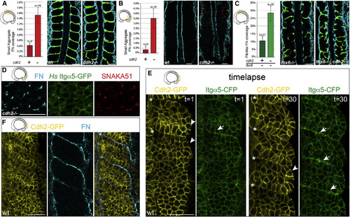
Cdh2 Represses Itgα5 Activation (A and B) cdh2-/- embryos produce significantly more ectopic small aggregates of FN matrix (red) in the mesenchyme of both (A) the anterior (p < 0.005 and t test) and (B) the posterior paraxial mesoderm (p < 0.02 and t test). (C) tbx6-/-;cdh2-/- embryos produce significantly more FN fibrils (green) in the anterior paraxial mesoderm mesenchyme than tbx6-/- controls (p < 0.01 and t test). The mean values ± SEM are shown. (D) Ectopic FN assembly, Itgα5 clustering, and adoption of the active Itgα5 conformation are observed in the PSM mesenchyme of cdh2 mutants (n = 38 embryos). (E) The first and last time points of a 90 min time-lapse (one frame every 3 min) of Cdh2-GFP and Itgα5-CFP localization during somite morphogenesis (see also Movie S3). The Cdh2-GFP levels decrease at somite boundaries (asterisks), while Itgα5-CFP levels increase (arrows) due to clustering. The Cdh2-GFP is also diminished along the surface of the paraxial mesoderm (arrowheads) (n = 4 time-lapses). (F) Cdh2-GFP levels anti-correlate with site of FN assembly along somite boundaries and the tissue surface (n = 8 embryos). The scale bars represent 40 µm. |
Reprinted from Developmental Cell, 34(1), Jülich, D., Cobb, G., Melo, A.M., McMillen, P., Lawton, A.K., Mochrie, S.G., Rhoades, E., Holley, S.A., Cross-Scale Integrin Regulation Organizes ECM and Tissue Topology, 33-44, Copyright (2015) with permission from Elsevier. Full text @ Dev. Cell