
FIGURE
Fig. S4
- ID
- ZDB-FIG-140324-47
- Publication
- Weber et al., 2013 - Characterization of light lesion paradigms and optical coherence tomography as tools to study adult retina regeneration in zebrafish
- Other Figures
- All Figure Page
- Back to All Figure Page
Fig. S4
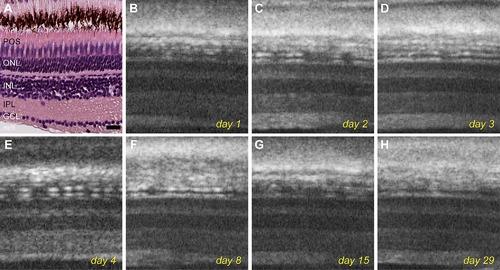
Live imaging of an untreated control fish over the course of 29 days. A: Histological staining of an untreated retina shows typical retinal layer structure. B–H: OCT images of the same fish acquired over 1 month shows no change in retinal structures. Scale bar represents 20 μm. |
Expression Data
Expression Detail
Antibody Labeling
Phenotype Data
Phenotype Detail
Acknowledgments
This image is the copyrighted work of the attributed author or publisher, and
ZFIN has permission only to display this image to its users.
Additional permissions should be obtained from the applicable author or publisher of the image.
Full text @ PLoS One