
Fig. 7
- ID
- ZDB-FIG-140324-39
- Publication
- Weber et al., 2013 - Characterization of light lesion paradigms and optical coherence tomography as tools to study adult retina regeneration in zebrafish
- Other Figures
- All Figure Page
- Back to All Figure Page
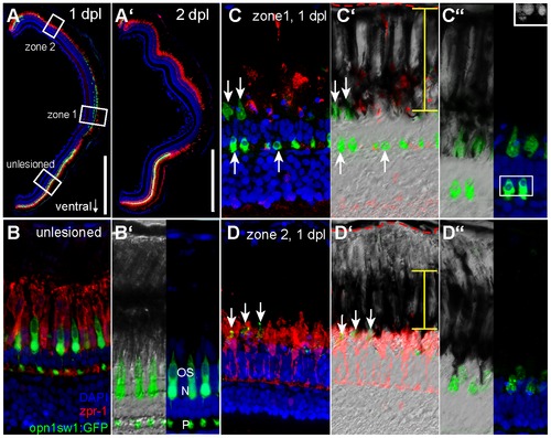
Differential removal of dying UV cones in zone 1 and 2. A: Overview image of a retinal section from an opn1sw1:GFP transgenic animal at 1 and 2 days post diffuse light lesion (A2). B: Unlesioned red/green double cones are labelled by zpr-1 (red) and UV cones by GFP. Nuclei were stained with DAPI (blue). B2: Close up of outer segments (OS), nucleus (N) and pedicle (P) is in the OPL. C: Central retina (zone 1) showing removal of zpr1+ cones while disrupted UV cones persist (arrow). C2: DIC image showing the distribution of pigmented granula in the RPE (yellow bar) relative to Bruch′s membrane (red dashed line). C3: Inset shows pyknotic nuclei in DAPI channel. D: Peripheral lesion with intact red/green cones but depleted UV cones. D2: DIC image showing condensed RPE pigments. D3: Co-localisation of pigmented processes with remaining GFP debris. ONL: Outer Nuclear Layer; RPE: Retina Pigmented Epithelium. Scale bars represent 500 μm. |