
Fig. 3
- ID
- ZDB-FIG-140324-37
- Publication
- Weber et al., 2013 - Characterization of light lesion paradigms and optical coherence tomography as tools to study adult retina regeneration in zebrafish
- Other Figures
- All Figure Page
- Back to All Figure Page
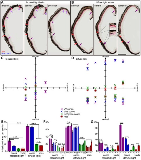
Extent of light lesioned areas in anterior-posterior and dorsal-ventral comparison. A, B: Double in situ hybridization against rhodopsin (red) to label rods and opn1sw1 (blue) to label UV cones. A: Focused light lesioned retinas in an anterior (left), central (middle) and posterior section (right). Central UV cone lesions are labeled with violet arrows, rod lesions are labeled with red arrows. Light purple and pink arrows indicate peripheral lesions of UV cones and rods, respectively. B: Diffuse light lesions analogous to A. Insets in the central section show the transition from healthy to lesioned areas. C: Blot of lesioned areas relative to their location in the retina. The average of dorsal and ventral lesion boundaries are blotted in the respective color code indicated in the legend. D: Lesioned areas analogous to C for diffuse light paradigm with additional far anterior and far posterior measure points. E: Comparison of average total damaged area in % of total retina length. F: Comparison of average anterior vs. posterior damaged area in % of total retina length. G: Comparison of average dorsal vs. ventral damaged area in % of 1/2 retina length. a: anterior, a2: far anterior, c: central, d: dorsal, p: posterior, p2: far posterior, r: rods; v: ventral. Column colors indicate cone type as in the legend (C). Error bars indicate standard error of the mean; *** for p-values <0.001; ** for p-values <0.01; * for p<0.05; n.s.: not significant; scale bar represents 400 μm. |