
FIGURE
Fig. S1
- ID
- ZDB-FIG-100121-24
- Publication
- LeClair et al., 2010 - Development and Regeneration of the Zebrafish Maxillary Barbel: A Novel Study System for Vertebrate Tissue Growth and Repair
- Other Figures
- All Figure Page
- Back to All Figure Page
Fig. S1
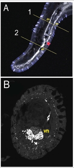
Abnormal regrowth of maxillary barbel nerve tracts A) Whole-mount immunohistochemistry of a regenerated barbel (7 dps). Nerves = white (acetylated tubulin). The red star indicates the amputation plane. Proximal to this level (1), there are two major nerves tracts, dorsal and ventral. Distal to this level (2), there is only one. dn = dorsal nerves; vn = ventral nerves. B) Section of the specimen at level 2, showing all nerve tracts displaced ventrally (vn). |
Expression Data
Expression Detail
Antibody Labeling
Phenotype Data
Phenotype Detail
Acknowledgments
This image is the copyrighted work of the attributed author or publisher, and
ZFIN has permission only to display this image to its users.
Additional permissions should be obtained from the applicable author or publisher of the image.
Full text @ PLoS One