
Fig. 5
- ID
- ZDB-FIG-100121-16
- Publication
- LeClair et al., 2010 - Development and Regeneration of the Zebrafish Maxillary Barbel: A Novel Study System for Vertebrate Tissue Growth and Repair
- Other Figures
- All Figure Page
- Back to All Figure Page
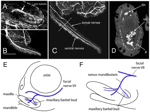
Innervation of the maxillary barbel. A) 75 μm barbel. In all panels, anterior is to the left. Whole-mount immunohistochemistry (anti-acetylated tubulin) shows a central tract of nerve fibers (arrow) within the early barbel bud (dotted white line). Smaller nerve projections are concentrated in the ventral half of the appendage. B) 200 μm barbel. Multiple fascicles of nerve fibers project distally, innervating the barbel′s ventral side and distal tip. No large tracts are visible dorsally. Scale bar = 100 μm. C) 1 mm barbel. Secondary nerve fibers appear within the dorsal half of the barbel. D) Section of an adult barbel at the approximate level shown by the dotted line in C. Innervation is visible as two deep nerve tracts (dn and vn) and a ring of sub-epithelial immunoreactive punctae (p). E,F) Schematic reconstructions of maxillary barbel bud innervation based on confocal tracing of whole-mount acetylated tubulin immunostaining in multiple zebrafish juveniles. F is an enlargement of the jaw region in E. |