
FIGURE
Fig. 7
- ID
- ZDB-FIG-100121-18
- Publication
- LeClair et al., 2010 - Development and Regeneration of the Zebrafish Maxillary Barbel: A Novel Study System for Vertebrate Tissue Growth and Repair
- Other Figures
- All Figure Page
- Back to All Figure Page
Fig. 7
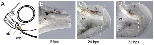
Proximal amputation of the maxillary barbel induces wound healing and blastema formation. A) Procedure for a unilateral “barbectomy”. The left maxillary barbel (mb) is amputated at the posterior margin of the maxilla. The contralateral barbel (not shown) is left as a non-surgical control. B–D) Fixed, unstained barbel regenerates collected immediately post surgery (B, 0 hps), or after 24 and 72 hours, respectively (C, D). Wound healing is followed by an accumulation of small, rounded mesenchymal cells underneath the epithelium (e) and around the central rod (cr). |
Expression Data
Expression Detail
Antibody Labeling
Phenotype Data
Phenotype Detail
Acknowledgments
This image is the copyrighted work of the attributed author or publisher, and
ZFIN has permission only to display this image to its users.
Additional permissions should be obtained from the applicable author or publisher of the image.
Full text @ PLoS One