
Fig. 3
- ID
- ZDB-FIG-100121-14
- Publication
- LeClair et al., 2010 - Development and Regeneration of the Zebrafish Maxillary Barbel: A Novel Study System for Vertebrate Tissue Growth and Repair
- Other Figures
- All Figure Page
- Back to All Figure Page
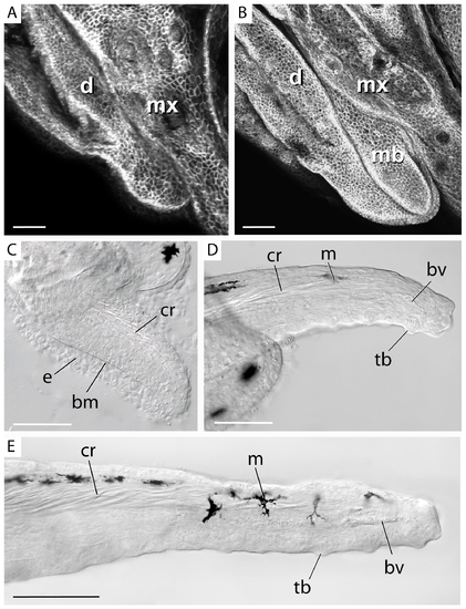
Early development of the maxillary barbel bud. A) Whole-mount confocal microscopy of the lower jaw of a juvenile membrane–GFP (mGFP) transgenic zebrafish (<10 mm standard length). Anterior is to the left. The maxillary barbel bud is not yet visible between the adjacent maxilla (mx) and dentary (d). All scale bars = 50 μm. B) Corresponding view of a slightly larger mGFP juvenile (10–12 mm SL). The first sign of the maxillary barbel (mb) is a rounded bud projecting caudoventrally. C) The early barbel bud has a thick outer epithelium (e) and a dense mesodermal core. The two layers are separated by a prominent basement membrane (bm). Fine strands of birefringent matrix accumulate dorsally where the central rod (cr) will form. D) As the barbel grows, the central rod becomes denser and projects farther distally. Isolated melanophores (m) appear along the dorsal epithelium. Ventrally, the blood vessel loop (bv) is forming. The ventral epithelium and distal tip carry numerous taste buds (tb). E) Later stages of barbel development involve expansion of the earlier structures. The central rod becomes longer and denser, the capillary loop extends, multiple melanophores become spaced along the dorsal surface, and numerous taste buds are added. The lymph vessel is not yet patent. bm = basement membrane; bv = blood vessel; cr = central rod; d = dentary; e = epithelium; m = melanophore; mb = maxillary barbel bud; ms = mesenchyme; mx = maxilla; o = orbit; tb = taste bud. |