
Fig. 8
- ID
- ZDB-FIG-100121-19
- Publication
- LeClair et al., 2010 - Development and Regeneration of the Zebrafish Maxillary Barbel: A Novel Study System for Vertebrate Tissue Growth and Repair
- Other Figures
- All Figure Page
- Back to All Figure Page
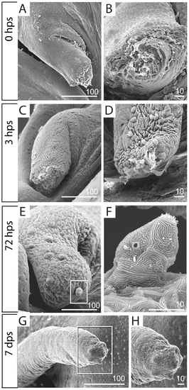
Scanning electron microscopy of early barbel regenerates. A, B) Immediately after amputation (zero hours post surgery, or 0 hps), the barbel stump is an open wound exposing the central core. A) Barbel stump in lateral view; proximal is to the left. B) End-on view of the same specimen. C, D) Two separate specimens collected at three hours post surgery (3 dps). The adjacent epithelium has closed the wound completely. Note the “purse string” lines of contraction within the epithelial sheet (C). Erythrocytes, dying cells, and matrix debris adhere to the distal surface (D). E,F) After 72 hours, the barbel stump swells distally, becoming bulbous (E). The tip epithelium carries newly differentiating taste bud hillocks, complete with protruding apical villi (F, magnified from E). G,H) By 7 days post surgery (7 dps), the maxillary barbel is a smaller version of the original appendage. Several millimeters long, it has a tapered distal end that carries a dense cluster of taste buds (H, magnified from G). |