
Fig. 12
- ID
- ZDB-FIG-100121-23
- Publication
- LeClair et al., 2010 - Development and Regeneration of the Zebrafish Maxillary Barbel: A Novel Study System for Vertebrate Tissue Growth and Repair
- Other Figures
- All Figure Page
- Back to All Figure Page
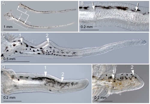
Repeated amputation can induce secondary regeneration. A) Two maxillary barbels regenerated from the same stump. The original barbel (not shown) was amputated at site 1 and the stump allowed to regenerate for one month. The resulting appendage (primary regenerate, top) was then amputated again slightly distal to the first amputation plane (site 2). A secondary regenerate (bottom) grew that was similar in size, shape and pigmentation to the primary regenerate. B) A magnification of the two surgical sites in A. Primary and secondary scars are visible approximately 0.5 mm apart. Note that the epithelial surface and melanophore patterning are largely normal. C) A secondary regenerate with more extreme scarring and swelling at the primary (1) and secondary (2) surgical sites. D–E) Failure of secondary regeneration. Secondary regenerates often failed to grow, elongating either slightly (D) or not at all (E) past the secondary surgical site (2). |