
Fig. 9
- ID
- ZDB-FIG-100121-20
- Publication
- LeClair et al., 2010 - Development and Regeneration of the Zebrafish Maxillary Barbel: A Novel Study System for Vertebrate Tissue Growth and Repair
- Other Figures
- All Figure Page
- Back to All Figure Page
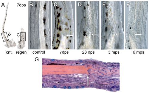
Internal scarring of maxillary barbel regenerates. A) Gross morphology of matched maxillary barbels (cntl = control; regen = regenerate) collected 7 days post surgery (dps). Note that the regenerate is thicker than the contralateral control and contains a central rod “stump.” B) Magnification of the control barbel in A. C) Magnification of the regenerated barbel in A. Note the absence of the central rod and the presence of wavy strands of matrix distal to the original amputation plane (arrow). The epithelial surface, pigment cell patterning and capillary loop are largely normal. D–F) Three regenerated barbels collected at 1–6 months post surgery (mps). All show disorganized mesodermal cores distal to the plane of section (arrow). G) Longitudinal histological section of a maxillary barbel regenerate (10 dps) showing disorganization of mesodermal cells and extracellular matrix distal to the amputation plane (arrow). Proximal is to the left. Hematoxylin/eosin stain. |