
Fig. 11
- ID
- ZDB-FIG-100121-22
- Publication
- LeClair et al., 2010 - Development and Regeneration of the Zebrafish Maxillary Barbel: A Novel Study System for Vertebrate Tissue Growth and Repair
- Other Figures
- All Figure Page
- Back to All Figure Page
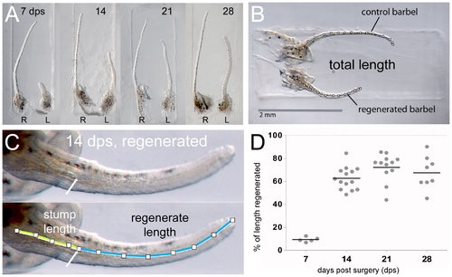
Barbel regeneration does not restore appendage length. A) Four matched pairs of maxillary barbels collected 7–28 days post surgery and embedded in the wells of a DNA electrophoresis gel. R = right barbel (unoperated control); L = left barbel (regenerate). All regenerates are shorter than the contralateral appendages. Each panel is shown at the same magnification. B) Measurement of total length (TL). The total length of each barbel (segmented line) was measured from the base of the central rod to the distal tip of the appendage. C) Measurement of stump length (SL) and regenerate length (RL). Within each regenerating barbel, the stump was measured from the base of the central rod to the amputation plane. The regenerate length was measured from the amputation plane to the distal tip. Stump length + regenerate length = total length of the regenerating barbel (SL + RL = TL). D) The regrowth of each regenerate was calculated as a percent of the control (% of length regenerated = (regenerate length/(control length – stump length))*100). Most lengthening occurred 7–14 days post surgery. Longer periods of regeneration (21–28 days) did not yield statistically significant differences in length. |