
Fig. S1
- ID
- ZDB-FIG-090223-24
- Publication
- Hinits et al., 2009 - Differential requirements for myogenic regulatory factors distinguish medial and lateral somitic, cranial and fin muscle fibre populations
- Other Figures
- All Figure Page
- Back to All Figure Page
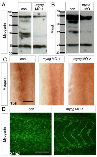
myog and myod MOs can specifically block translation of their mRNAs. (A,B) Western blot of protein extracts from control (A,B) and myog MO-1 (A) or myod MO (B), showing absence of specific bands at ∼37 kDa from the MO lanes. Myod immunohistochemical stain is also ablated (Hammond et al., 2007) #8381. (C) Immunohistochemistry with Myogenin antibody (brown nuclei) of myog MO-1-injected, myog MO-2-injected and control 15 ss embryos. Dorsal flatmounts of one side of anterior somites, anterior to top. (D) Confocal stacks of tail somites of 24 hpf control and myog MO-1-injected embryos after immunodetection of Myogenin (green nuclei). Lateral view, anterior to left. myog morphants lack the nuclear Myogenin reactivity of control embryos. |