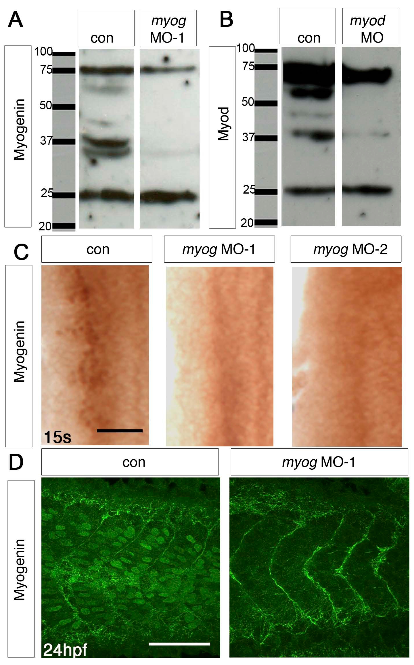
Fig. S1 myog and myod MOs can specifically block translation of their mRNAs. (A,B) Western blot of protein extracts from control (A,B) and myog MO-1 (A) or myod MO (B), showing absence of specific bands at ∼37 kDa from the MO lanes. Myod immunohistochemical stain is also ablated (Hammond et al., 2007) #8381. (C) Immunohistochemistry with Myogenin antibody (brown nuclei) of myog MO-1-injected, myog MO-2-injected and control 15 ss embryos. Dorsal flatmounts of one side of anterior somites, anterior to top. (D) Confocal stacks of tail somites of 24 hpf control and myog MO-1-injected embryos after immunodetection of Myogenin (green nuclei). Lateral view, anterior to left. myog morphants lack the nuclear Myogenin reactivity of control embryos.
Image
Figure Caption
Acknowledgments
This image is the copyrighted work of the attributed author or publisher, and
ZFIN has permission only to display this image to its users.
Additional permissions should be obtained from the applicable author or publisher of the image.
Full text @ Development

