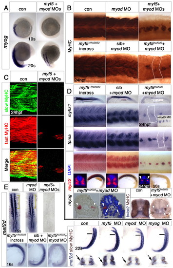
Myf5 or Myod is required for medial fast myogenesis. In situ hybridisation for myog, myhz1, tpma, mylz2 or mef2d or immunohistochemistry for the indicated myosins in wild-type or myf5+/hu2022 incross zebrafish injected with the indicated MOs. Lateral views, anterior to left unless otherwise stated. (A) myog expression was delayed in embryos injected with myf5+myod MOs. Dorsolateral views, anterior to top. (B) MyHC was reduced in both epaxial and hypaxial domains in myod MO embryos, grossly reduced in double myf5+myod morphants and further diminished in myf5hu2022 mutants injected with myod MO. Note the complete absence of residual cells in some individual somites (dotted outline). (C) Residual differentiated muscle in myf5+myod morphants expressed fast but not slow MyHC at 24 hpf. Lateral view of confocal stack of somites 9-10. (D) Fast muscle markers were indistinguishable in 24 hpf myf5hu2022 homozygous mutants and their siblings (left column), reduced by myod MO in siblings (middle) and almost eliminated by myod MO in myf5hu2022 mutants (right). Inset illustrates that myf5 MO does not worsen the myod MO plus myf5hu2022 phenotype. mylz2 mRNA accumulation (red, arrowheads) and myog downregulation (blue) are delayed at ∼20 ss in fast muscle. (Bottom panels) Transverse sections of double knockdowns show residual fast muscle (arrowheads) immediately lateral to adaxial and/or twist2-expressing sclerotomal cells (arrows). (E) (Left, top) mef2d mRNA initiates normally in pbcs in the myod morphant, but is restricted to the medial region of rostral somites (yellow brackets). In myf5+myod MOs, mef2d mRNA is absent. Dorsal flatmounts, anterior to top. (Left, bottom) Lack of both myf5 and myod ablates mef2d expression. Lateral views, anterior to top. (Right) By 22 ss, transverse sections (at the level of the bar on lateral flatmounts) show reduction of mef2d mRNA (purple) in myod or myog, but not in myf5, single morphants. Mef2 position is shown relative to slow fibres (brown). Note the expression of mef2d lateral to migrating slow fibres in the control and in myf5 and myog, but not myod, morphants (arrows).
|
