FIGURE SUMMARY
Title

Identification of a pharyngeal mucosal lymphoid organ in zebrafish and other teleosts: Tonsils in fish?

Authors
Resseguier, J., Nguyen-Chi, M., Wohlmann, J., Rigaudeau, D., Salinas, I., Oehlers, S.H., Wiegertjes, G.F., Johansen, F.E., Qiao, S.W., Koppang, E.O., Verrier, B., Boudinot, P., Griffiths, G.
Source
Full text @ Sci Adv

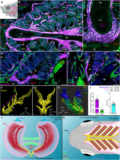
General organization and localization of NELO in zebrafish

(A) Scheme illustrating the different orientations of the NELO images acquired from 30-μm whole adult zebrafish head cryosections, and the position of the thymus (blue), pharynx (green), and gills (red). (B to E) NELO (red arrowheads) wraps around the urohyal bone (B and C) and extends along the sub-pharyngeal isthmus toward the posterior end of the gill chambers (D). NELO is connected to the ILTs [(B), yellow stars], the ALTs [(E), cyan stars], and the cavo-branchial epithelium [(A), cyan arrowheads]. NELO is close to the gills afferent arteries [(B), cyan arrows] and other endothelial vessels [(C), orange arrows]. (F and G) NELO 3D reconstruction obtained by serial confocal tomography of a ZAP70-labeled wholemount head of zebrafish (15 wpf) and its segmentation into four anatomic regions: the anterior area wrapped around the urohyal bone (cyan arrowhead), antler-like protrusions (magenta arrowheads), the core (blue arrow), and the posterior end (green arrowheads). (H) Three-dimensional reconstruction of NELO (yellow), ALTs (cyan), thymus lobes (blue), and the ventral extremity of gill arches (green). (I) Volumes of the 3D reconstructed lymphoid structures, a fraction of the volume occupied by T/NK cells in NELO and the spleen, and the average volume of a single T/NK cell. (J and K) Illustrations of NELO’s localization as observed from the front (J) or from below (K). Illustrations made by Ella Maru studio and K. Zulkefli. Annotations: Aa, afferent artery; ALT, amphibranchial lymphoid tissue; Bc, branchial cavity; C, cartilage; Cbe, cavo-branchial epithelium; Cvs, central venous sinus; Ea, efferent artery; Ga, gill arch; ILT, interbranchial lymphoid tissue; La, lamellae; M, muscles; NELO, Nemausean lymphoid organ; S, septum; Spi, sub-pharyngeal isthmus; Td, tendon; Tf, thyroid follicle; Uh, urohyal bone; Va, ventral aorta. Scale bars, 150 (H), 100 (B, and E to G), 50 (D), and 40 μm (C).

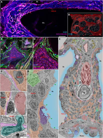
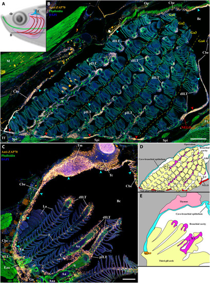
Detailed structural organization of the adult zebrafish NELO.

(A) Cryosection labeled with anti-cytokeratin antibodies (magenta hot) revealing a network of reticulated epithelial cells within (yellow arrowheads) and bordering NELO (red arrowheads). (B) Three-dimensional reconstruction illustrating the network of reticulated epithelial cells in red. (C) Three-dimensional imaging of a cryosection displaying NELO from a fli:GFP zebrafish, in which endothelial vessels are fluorescent (green). Numerous vessels are wrapped around NELO (cyan arrows). (D) Optical section from (C) highlighting cuboidal-shaped endothelial cells (cyan arrowheads). (E to H) Ultrastructure map of a 9 wpf zebrafish NELO transversally sectioned at the urohyal bone acquired by transmission electron microscopy. Several structures have been highlighted: reticulated epithelial cells (orange), mucous cells (dark blue), water (light blue), ionocytes (purple), endothelial vessels (burgundy red), basement membrane (pink), neutrophils (green), basophils/mast cells (yellow), tenocytes (dark blue-green), and pavement cells (green arrowheads). (F) to (H) represent zoomed areas from (E). (I) Cell (dark blue-green) observed across the basement membrane (pink) separating NELO from the surrounding connective tissue. Annotations: Bm, basement membrane; Gi, Gills; Sk, Skin. Scale bars, 30 μm (C), 20 μm (A), 10 μm (D), 4 μm (E), 3 μm (B), 1 μm (G, H, and I), and 500 nm (F).

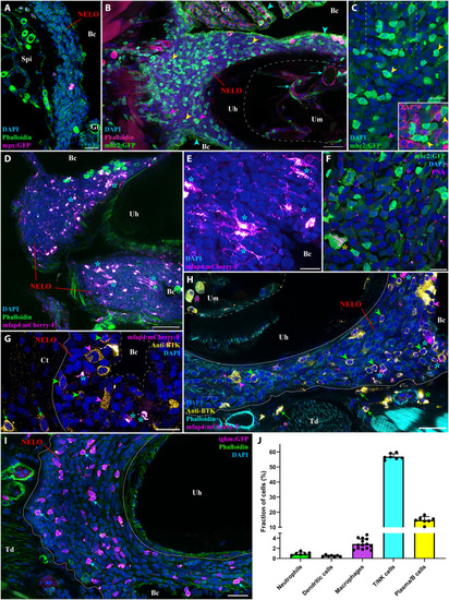
NELO immune cell populations

(A) Adult mpx:GFP zebrafish NELO cryosection, in which neutrophils are fluorescent (magenta hot). (B) Adult mhc2:GFP zebrafish cryosection, in which mhc2-expressing cells are fluorescent (green). The expression of MHC2 is observed in the epithelial cells (cyan arrowheads), large cells inside NELO (magenta arrowheads), and within the marrow of the urohyal bone (cyan arrows). (C) Zoom inside NELO of a mhc2:GFP fish highlighting large (magenta arrowheads) and small (yellow arrowheads) positive cells. The latter being negative for anti-ZAP70 labeling (cherry). (D and E) Adult mfap4:mCherry-F zebrafish NELO cryosection in which macrophages are fluorescent (magenta hot, cyan stars). (F) Cryosection of an adult mhc2:GFP zebrafish NELO stained with peanut agglutinin lectin (magenta hot) to reveal dendritic cell (yellow star). (G and H) Anti-Bruton’s tyrosine kinase (BTK) labeling (yellow hot) of mfap4:mCherry-F adult zebrafish cryosections revealed macrophages (magenta hot, cyan stars), BTK-positive B cells (green arrowheads) and BTK-positive plasma cells (magenta arrowheads) in NELO. BTK-positive cells were also found in the connective tissue surrounding NELO (green arrows), around endothelial structures (green stars), and within the marrow of the urohyal bone (magenta star). (I) The presence of B cells in NELO was confirmed using ighm:GFP zebrafish, in which a subtype of B cells that express immunoglobulin M is fluorescent (magenta hot, cyan arrows). (J) Quantification of the different immune cell populations found in NELO counted from at least seven single-cell layer images originating from at least three fish. Annotation: Um, Urohyal marrow. Scale bars, 50 (D), 30 (B), 20 (A, H, and I), and 10 (C and E to G).

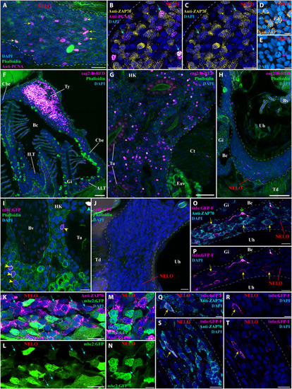
Investigation of immune function molecular markers in NELO.

(A) NELO cryosection labeled with anti-PCNA antibody (magenta hot) to reveal proliferating cells (yellow arrows). (B and C) Cryosection co-labeled with anti-PCNA (magenta hot) and anti-ZAP70 (yellow hot) to reveal the presence of proliferative T/NK cells in NELO (cyan arrows). (D and E) The presence of proliferative T/NK cells in NELO was confirmed by the presence of ZAP70-positive cells (orange hot) displaying mitotic figures (magenta arrow). (F to H) Cryosections from rag2:DsRED zebrafish in which cells undergoing V(D)J recombination are fluorescent (magenta hot). Whereas numerous positive cells are found in the thymus (F) and the head-kidney (G), which are known sites of V(D)J recombination for T and B cells, almost none were observed in NELO (H), the ILTs and the ALTs (F). (I and J) Cryosections from cd41:GFP zebrafish, in which thrombocytes (cyan arrowhead) are brightly fluorescent and hematopoietic stem cells are faintly fluorescent (yellow arrowhead) (magenta hot). In contrast to the expected localization of hematopoietic stem cells in the kidney (I), none were observed in NELO (J). (K to N) Cryosections from mhc2:GFP zebrafish (green) labeled with anti-ZAP70 (magenta hot) revealed the presence of mhc2-expressing T/NK cells (cyan arrows), a feature of activated T/NK cells. (O to T) Cryosections from tnfα:GFP zebrafish NELO, in which cells expressing the immune effector molecule tumor necrosis factor–α (TNF-α) are fluorescent (magenta hot), labeled with anti-ZAP70 (cyan). Annotations: Bv, blood vessel; Ct, connective tissue; Eav, endothelium anastomotic vessels; HK, head-kidney; Tu, tubule; Ty, thymus. Scale bars, 50 (F to H), 20 (A), 10 (B, C, I to L, O, P, S, and T), and 5 μm (D, E, M, N, Q, and R).

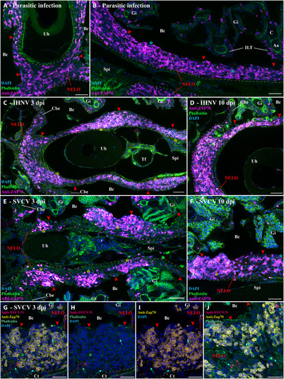
Structural response of NELO to viral and parasitic infections.

(A and B) Cryosections displaying NELO (red arrowheads) in adult zebrafish naturally coinfected with three parasite diseases (P. neurophilia, P. tomentosa, and M. streisingeri) labeled with anti-ZAP70 antibody (magenta hot). The distribution of ZAP70-positive cells in NELO appears more heterogeneous than in uninfected fish. In addition, some the labeled cells formed small clusters (cyan stars). (C and D) Cryosections from adult zebrafish bath-infected for 24 hours with IHNV. After the apparition of more prominent aggregations of ZAP70-positive cells at 3 dpi (yellow stars) (C), the distribution of T/NK cells reverted to a more homogeneous state by 10 dpi (D). (E and F) Cryosections from adult zebrafish bath-infected for 24 hours with SVCV. NELO displayed notable aggregations of T/NK cells into distinct clusters at 3 dpi (yellow stars) (E). A week later, NELO displayed both large (yellow star) and small clusters (cyan star) of ZAP70-positive cells (F). (G to J) Cryosections from zebrafish 3 day after SVCV infection co-labeled with anti-ZAP70 antibody (yellow) and anti-SVCV-N antibody (cherry), revealing cells loaded with viral material (cyan arrows) neighboring large clusters of ZAP70-positive cells. Annotations: dpi, day postinfection; IHNV, infectious hematopoietic necrosis virus; SVCV, spring viremia of carp virus. Scale bars, 50 (E), 30 (A to D and F), and 20 μm (G to J).

NELO as part of a larger lymphoid network.

(A) Scheme illustrating the localization of the branchial cavity images acquired from 30-μm whole adult zebrafish head cryosections labeled with anti-ZAP70 antibody (orange hot) to reveal T/NK cells (B and C). NELO (red arrowheads) is part of the lymphoid network propagated by the ZAP70-positive cell–rich cavo-branchial epithelium (cyan arrowheads) and which connects all the lymphoid structures of the branchial cavity. (D) Scheme describing the different anatomical regions displayed by the coronal cryosection presented in (B). (E) Scheme describing the different anatomical regions displayed by the transversal cryosection presented in (C). Annotations: Aaa, afferent arch artery; dILT, distal interbranchial lymphoid tissue; Eaa, efferent arch artery; Ft, fat tissue; Op, operculum; pILT, proximal interbranchial lymphoid tissue; Tc, thymus cortex; Tm, thymus medulla. Scale bars, 200 (B) and 100 μm (C).

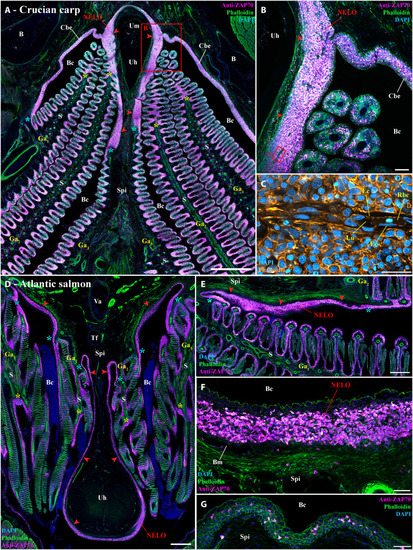
NELO and its cohesion with ILTs and ALTs in other teleost fish species.

(A and B) Cryosection from wild crucian carp labeled with anti-ZAP70 antibody (magenta hot). This larger representative of the cyprinids than zebrafish also has NELO (red arrowheads), ILTs (yellow stars), and ALTs (cyan stars), which are all interconnected by a clear lymphoid network. (C) Zoom from (B) in which phalloidin (orange hot) and DAPI (cyan hot) staining revealed the presence of endothelial vessels containing red blood cells. Transversal (D) and coronal (E) cryosections of an adult Atlantic salmon (1.5 kg) labeled with anti-ZAP70 (magenta hot) displaying NELO (red arrowheads), ILTs (yellow stars), and ALTs (cyan stars). As for cyprinids, NELO (red arrowheads) of this representative of the salmonid family is located at the ventral convergence of gill arches, interconnected with gills lymphoid aggregates, and closely associated with the urohyal bone. It is particularly enriched in ZAP70-positive cells (F). In contrast, the mucosal epithelium lining the region directly anterior to the urohyal bone only displayed scarce T/NK cell (G). Annotations: B, bone; Ec, endothelial cell; Lu, lumen; Rbc, red blood cell. Scale bars, 1000 (A), 500 (D), 400 (E), 100 (B), 50 (F and G), and 20 μm (C).

Acknowledgments
This image is the copyrighted work of the attributed author or publisher, and ZFIN has permission only to display this image to its users. Additional permissions should be obtained from the applicable author or publisher of the image. Full text @ Sci Adv