IMAGE

Fig. 2.

ID
ZDB-IMAGE-231104-2
Source
Figures for Resseguier et al., 2023
Image
Figure Caption

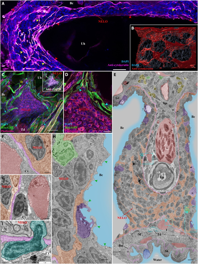
Fig. 2. Detailed structural organization of the adult zebrafish NELO.

(A) Cryosection labeled with anti-cytokeratin antibodies (magenta hot) revealing a network of reticulated epithelial cells within (yellow arrowheads) and bordering NELO (red arrowheads). (B) Three-dimensional reconstruction illustrating the network of reticulated epithelial cells in red. (C) Three-dimensional imaging of a cryosection displaying NELO from a fli:GFP zebrafish, in which endothelial vessels are fluorescent (green). Numerous vessels are wrapped around NELO (cyan arrows). (D) Optical section from (C) highlighting cuboidal-shaped endothelial cells (cyan arrowheads). (E to H) Ultrastructure map of a 9 wpf zebrafish NELO transversally sectioned at the urohyal bone acquired by transmission electron microscopy. Several structures have been highlighted: reticulated epithelial cells (orange), mucous cells (dark blue), water (light blue), ionocytes (purple), endothelial vessels (burgundy red), basement membrane (pink), neutrophils (green), basophils/mast cells (yellow), tenocytes (dark blue-green), and pavement cells (green arrowheads). (F) to (H) represent zoomed areas from (E). (I) Cell (dark blue-green) observed across the basement membrane (pink) separating NELO from the surrounding connective tissue. Annotations: Bm, basement membrane; Gi, Gills; Sk, Skin. Scale bars, 30 μm (C), 20 μm (A), 10 μm (D), 4 μm (E), 3 μm (B), 1 μm (G, H, and I), and 500 nm (F).

Acknowledgments
This image is the copyrighted work of the attributed author or publisher, and ZFIN has permission only to display this image to its users. Additional permissions should be obtained from the applicable author or publisher of the image. Full text @ Sci Adv