|
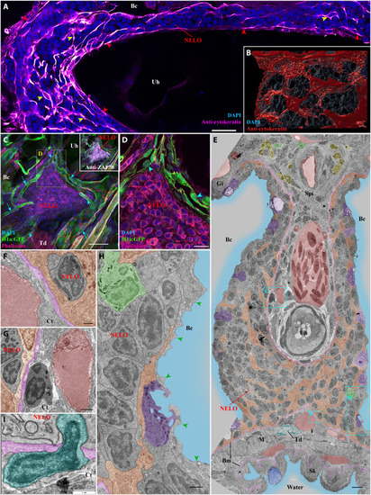
Detailed structural organization of the adult zebrafish NELO. (A) Cryosection labeled with anti-cytokeratin antibodies (magenta hot) revealing a network of reticulated epithelial cells within (yellow arrowheads) and bordering NELO (red arrowheads). (B) Three-dimensional reconstruction illustrating the network of reticulated epithelial cells in red. (C) Three-dimensional imaging of a cryosection displaying NELO from a fli:GFP zebrafish, in which endothelial vessels are fluorescent (green). Numerous vessels are wrapped around NELO (cyan arrows). (D) Optical section from (C) highlighting cuboidal-shaped endothelial cells (cyan arrowheads). (E to H) Ultrastructure map of a 9 wpf zebrafish NELO transversally sectioned at the urohyal bone acquired by transmission electron microscopy. Several structures have been highlighted: reticulated epithelial cells (orange), mucous cells (dark blue), water (light blue), ionocytes (purple), endothelial vessels (burgundy red), basement membrane (pink), neutrophils (green), basophils/mast cells (yellow), tenocytes (dark blue-green), and pavement cells (green arrowheads). (F) to (H) represent zoomed areas from (E). (I) Cell (dark blue-green) observed across the basement membrane (pink) separating NELO from the surrounding connective tissue. Annotations: Bm, basement membrane; Gi, Gills; Sk, Skin. Scale bars, 30 μm (C), 20 μm (A), 10 μm (D), 4 μm (E), 3 μm (B), 1 μm (G, H, and I), and 500 nm (F).
|