|
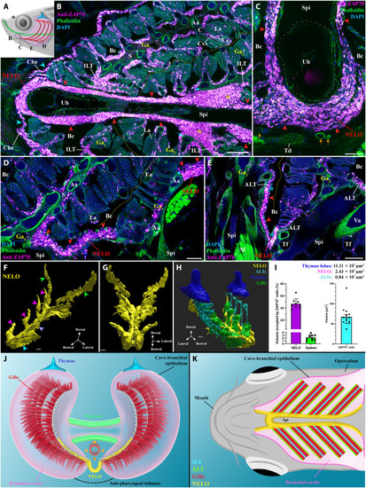
General organization and localization of NELO in zebrafish (A) Scheme illustrating the different orientations of the NELO images acquired from 30-μm whole adult zebrafish head cryosections, and the position of the thymus (blue), pharynx (green), and gills (red). (B to E) NELO (red arrowheads) wraps around the urohyal bone (B and C) and extends along the sub-pharyngeal isthmus toward the posterior end of the gill chambers (D). NELO is connected to the ILTs [(B), yellow stars], the ALTs [(E), cyan stars], and the cavo-branchial epithelium [(A), cyan arrowheads]. NELO is close to the gills afferent arteries [(B), cyan arrows] and other endothelial vessels [(C), orange arrows]. (F and G) NELO 3D reconstruction obtained by serial confocal tomography of a ZAP70-labeled wholemount head of zebrafish (15 wpf) and its segmentation into four anatomic regions: the anterior area wrapped around the urohyal bone (cyan arrowhead), antler-like protrusions (magenta arrowheads), the core (blue arrow), and the posterior end (green arrowheads). (H) Three-dimensional reconstruction of NELO (yellow), ALTs (cyan), thymus lobes (blue), and the ventral extremity of gill arches (green). (I) Volumes of the 3D reconstructed lymphoid structures, a fraction of the volume occupied by T/NK cells in NELO and the spleen, and the average volume of a single T/NK cell. (J and K) Illustrations of NELO’s localization as observed from the front (J) or from below (K). Illustrations made by Ella Maru studio and K. Zulkefli. Annotations: Aa, afferent artery; ALT, amphibranchial lymphoid tissue; Bc, branchial cavity; C, cartilage; Cbe, cavo-branchial epithelium; Cvs, central venous sinus; Ea, efferent artery; Ga, gill arch; ILT, interbranchial lymphoid tissue; La, lamellae; M, muscles; NELO, Nemausean lymphoid organ; S, septum; Spi, sub-pharyngeal isthmus; Td, tendon; Tf, thyroid follicle; Uh, urohyal bone; Va, ventral aorta. Scale bars, 150 (H), 100 (B, and E to G), 50 (D), and 40 μm (C).
|