|
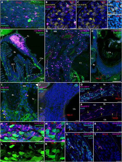
Investigation of immune function molecular markers in NELO. (A) NELO cryosection labeled with anti-PCNA antibody (magenta hot) to reveal proliferating cells (yellow arrows). (B and C) Cryosection co-labeled with anti-PCNA (magenta hot) and anti-ZAP70 (yellow hot) to reveal the presence of proliferative T/NK cells in NELO (cyan arrows). (D and E) The presence of proliferative T/NK cells in NELO was confirmed by the presence of ZAP70-positive cells (orange hot) displaying mitotic figures (magenta arrow). (F to H) Cryosections from rag2:DsRED zebrafish in which cells undergoing V(D)J recombination are fluorescent (magenta hot). Whereas numerous positive cells are found in the thymus (F) and the head-kidney (G), which are known sites of V(D)J recombination for T and B cells, almost none were observed in NELO (H), the ILTs and the ALTs (F). (I and J) Cryosections from cd41:GFP zebrafish, in which thrombocytes (cyan arrowhead) are brightly fluorescent and hematopoietic stem cells are faintly fluorescent (yellow arrowhead) (magenta hot). In contrast to the expected localization of hematopoietic stem cells in the kidney (I), none were observed in NELO (J). (K to N) Cryosections from mhc2:GFP zebrafish (green) labeled with anti-ZAP70 (magenta hot) revealed the presence of mhc2-expressing T/NK cells (cyan arrows), a feature of activated T/NK cells. (O to T) Cryosections from tnfα:GFP zebrafish NELO, in which cells expressing the immune effector molecule tumor necrosis factor–α (TNF-α) are fluorescent (magenta hot), labeled with anti-ZAP70 (cyan). Annotations: Bv, blood vessel; Ct, connective tissue; Eav, endothelium anastomotic vessels; HK, head-kidney; Tu, tubule; Ty, thymus. Scale bars, 50 (F to H), 20 (A), 10 (B, C, I to L, O, P, S, and T), and 5 μm (D, E, M, N, Q, and R).
|