IMAGE

Fig. 6.

ID
ZDB-IMAGE-231104-6
Source
Figures for Resseguier et al., 2023
Image
Figure Caption

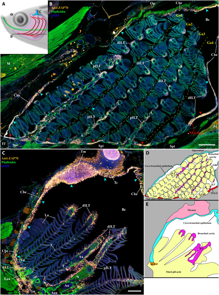
Fig. 6. NELO as part of a larger lymphoid network.

(A) Scheme illustrating the localization of the branchial cavity images acquired from 30-μm whole adult zebrafish head cryosections labeled with anti-ZAP70 antibody (orange hot) to reveal T/NK cells (B and C). NELO (red arrowheads) is part of the lymphoid network propagated by the ZAP70-positive cell–rich cavo-branchial epithelium (cyan arrowheads) and which connects all the lymphoid structures of the branchial cavity. (D) Scheme describing the different anatomical regions displayed by the coronal cryosection presented in (B). (E) Scheme describing the different anatomical regions displayed by the transversal cryosection presented in (C). Annotations: Aaa, afferent arch artery; dILT, distal interbranchial lymphoid tissue; Eaa, efferent arch artery; Ft, fat tissue; Op, operculum; pILT, proximal interbranchial lymphoid tissue; Tc, thymus cortex; Tm, thymus medulla. Scale bars, 200 (B) and 100 μm (C).

Acknowledgments
This image is the copyrighted work of the attributed author or publisher, and ZFIN has permission only to display this image to its users. Additional permissions should be obtained from the applicable author or publisher of the image. Full text @ Sci Adv