
- Title
-
The conundrum of pharyngeal teeth origin: the role of germ layers, pouches, and gill slits
- Authors
- Huysseune, A., Cerny, R., Witten, P.E.
- Source
- Full text @ Biol. Rev. Camb. Philos. Soc.
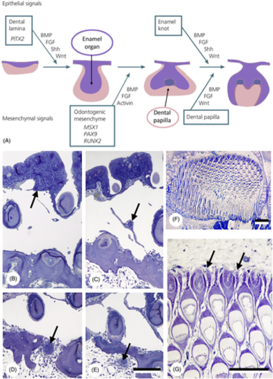
Epithelial primacy in tooth formation. (A) Schematic representation of tooth formation in mammals and transcription factors and signalling molecules involved (modified from Koch, Thesleff & Kreiborg, 2017); purple: epithelium/enamel organ; pink: mesenchyme/dental papilla. BMP, bone morphogenetic protein; FGF, fibroblast growth factor; Shh, sonic hedgehog. (B–E) Intramedullary tooth formation in the lower pharyngeal jaw of the cichlid Hemichromis bimaculatus, 28 mm standard length (SL). Successive cross sections through the epithelial strand (arrow) giving rise to the enamel organ of the replacement tooth, showing its origin (B), its course across the large vascular cavity (C), its penetration through the jaw bone (D) and its distal end within the medullary cavity of the jaw bone (E). Only few mesenchymal cells have condensed around the distal end. Total length of the epithelial strand is approximately 300 μm. Scale bar for B–E = 100 μm. (F, G) Section through the left premaxilla of a scraping loricariid catfish, Ancistrus cf. triradiatus, of 33.5 mm SL. Note virtual absence of mesenchymal cells at the distal end of each successional lamina (arrows). Scale bars: F = 100 μm; G = 50 μm. |
Schematic representation of primary mouth formation in vertebrates [from Soukup et al., 2013, with permission from the authors and publisher]. Yellow, endoderm; blue, ectoderm. |
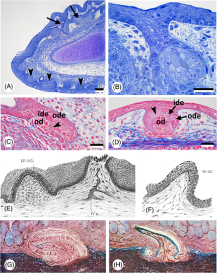
Oropharyngeal epithelium, teeth, and dermal denticles in chondrichthyans. (A) Scyliorhinus canicula; sagittal section of the lower jaw of a 85 mm embryo, showing skin (= dermal) denticles (arrowheads), and the dental lamina with developing teeth (arrows). Scale bar = 100 μm. (B) Scyliorhinus canicula; larger magnification of the early dental lamina with a developing tooth in a 85 mm embryo. Scale bar = 20 μm. (C, D) Scyliorhinus canicula; sagittal section of a tooth anlage in the lower jaw with the invading dental lamina (C) compared to the anlage of a dermal denticle (D) in the same 50 mm embryo. Outer (ode) and inner dental epithelium (ide), the basal lamina of the enamel organ (arrowhead) and the mesenchymal condensation that will give rise to the odontoblasts (od) characterise the anlagen of teeth and dermal denticles. Scale bars = 25 μm. (E, F) Squalus acanthias; two stages of development of a ‘placoid scale’ (from Cook & Neal, 1921). n'v. = nerve; pl. sc. = placoid scale. (G, H) Mustelus sp.; two stages of development of a ‘mucous membrane denticle’ (from Peyer, 1968). |
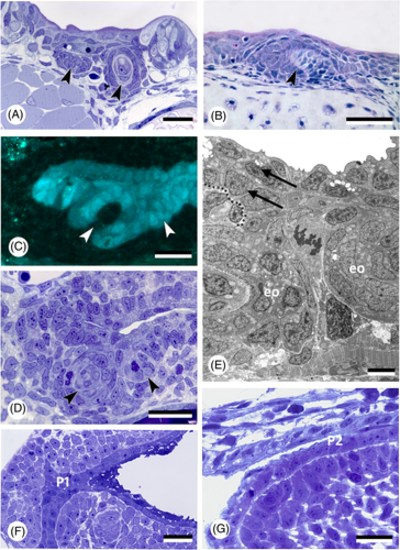
Oropharyngeal epithelium and teeth in actinopterygians. (A) Polypterus senegalus; pharyngeal epithelium and forming tooth germs (arrowheads) in an 18 mm larva. Scale bar = 20 μm. (B) Acipenser ruthenus; tooth germ (arrowhead) in the buccopharyngeal epithelium in a 15 mm larva. Scale bar = 50 μm. (C, D) Danio rerio, cross section of a 56 h post-fertilisation (hpf) embryo at the level of the first forming teeth (arrowheads), shown in a transgenic (sox17:egfp) line (C), and corresponding semithin, toluidine blue-stained section of a wildtype fish (D). Scale bars = 20 μm. (E) Danio rerio, transmission electron microscopy (TEM) image of the bilayered pharyngeal epithelium (two arrows, delimited by dotted line) of a 72 hpf embryo at the level of the first two teeth (eo, enamel organ). Scale bar = 5 μm. (F) Hemichromis bimaculatus, cross section of the oropharynx of a 1 day post-hatching (dph) (4.0 mm total length) specimen at the level of pouch 1 (P1). Scale bar = 20 μm. (G) Oncorhynchus tshawytscha, sagittal section of the pharyngeal epithelium in an unhatched embryo 17 days post-fertilisation (dpf), at the level of the (open) pouch 2 (P2). Scale bar = 20 μm. |
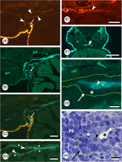
Germ layer contribution to oropharyngeal epithelium and teeth in zebrafish, Danio rerio. (A–C) Sagittal section of a double-transgenic (krt4:tomatoCAAX;sox17:egfp) 55 h post-fertilisation (hpf) embryo showing periderm having partially invaded pouch 2 (intense orange), and connecting to periderm-like cells investing the oropharynx (red, indicated by white arrowheads, shown also in E); boundary indicated by a small white arrow; both populations do not show further mixing. Micrographs are shown in the red (A) and green channel (B), and as overlay (C). Dotted line in C marks the outline of pouch 2. Scale bar for A–C = 20 μm. (D) Magnification of the pharynx lining shown in C; periderm-like cells (arrowheads) express both krt4 and sox17 and cover endoderm, in which sox17 is downregulated (asterisks). Scale bar = 20 μm. (E) Sagittal section of a transgenic (krt4:tomatoCAAX) 55 hpf embryo showing periderm-like layer (arrowhead). A lumen has appeared rostrally between the two periderm-like layers (asterisk). Scale bar = 20 μm. (F) Cross section of a 60 hpf Tg(krt4:gfp) embryo at the level of the forming pharyngeal dentition; periderm-like layer indicated by an arrowhead. Scale bar = 100 μm. (G) Magnification of F, showing the placode of the first pharyngeal tooth (arrow). This develops from the unlabelled basal (endodermal) layer (large asterisk), while being covered by a krt4-positive superficial layer (arrowhead). A dotted line delimits the pharyngeal epithelium; a lumen starts to appear between the two layers of periderm-like cells (small asterisk). Scale bar = 10 μm. (H) Toluidine blue-stained semithin cross section, showing placode stage of first pharyngeal tooth formation corresponding to G (tooth placode indicated by a dotted outline). Note superficial periderm-like layer (arrowhead) with forming pharyngeal lumen (asterisk). Laterally, the pouch (pouch 6, P6) is still bilayered. Scale bar = 10 μm. |
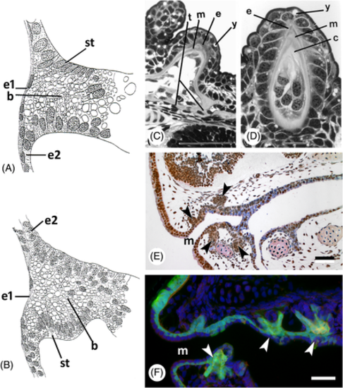
Germ layer contribution to oropharyngeal epithelium and teeth in lungfish and amphibians. (A) Lepidosiren paradoxa; drawing of a sagittal section of a stage 30 specimen (after Kerr, 1903); drawing mirrored to show anterior to the left. b, yolk-laden cells in the buccal cavity; e1, superficial ectodermal layer; e2, basal ectodermal layer; st, lining epithelium of buccal cavity arising in situ. (B) Ambystoma mexicanum; drawing of a similar section to A, embryo of 7.5 mm length (after Kerr, 1903; drawing mirrored to show anterior to the left). (C, D) Neoceratodus forsteri, cusp of pterygopalatine tooth plate [from Kemp, 2002a, with permission from the author and publisher]. c, circumdenteonal dentine; e, enamel; m, mantle dentine; t, trabeculae; y, yolk remnants. Scale bars: C = 100 μm; D = 20 μm. (E) Ambystoma mexicanum; sagittal section of a stage 43 embryo; compare with F. Tooth buds indicated by arrowheads. m, mouth opening. Scale bar = 50 μm. (F) Ambystoma mexicanum; transgenic embryo of stage 43 with green fluorescent protein (GFP) labelling in the ectoderm [modified from Soukup et al., 2008, courtesy of the authors]. Arrowheads: tooth buds arising in ectoderm; m, mouth opening. Scale bar = 50 μm. |
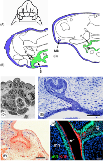
Germ layer contribution to oropharyngeal epithelium and teeth in amphibians and amniotes. (A) Ambystoma punctatum, extirpation of the ectoderm of a stage 24 embryo, ventral view (redrawn after Adams, 1924). (B) Ambystoma punctatum; sagittal section of a stage 39 embryo after extirpation of the ectoderm; teeth in the ectoderm (blue) are reduced or absent, while endoderm (green) has given rise to splenial (S) and palatine (P) tooth germs (red); FG, foregut (adapted after Adams, 1924). (C) Ambystoma punctatum; sagittal section of a stage 45 embryo showing a maxillary bulb (MB) separated from any connection with the foregut; no contact between ectoderm (blue) and endoderm (green), yet a palatine tooth (P) is present (red); FG, foregut (adapted after Adams, 1924). (D) Pleurodeles waltl, first-generation tooth, stage 36. dp, dental papilla; ide, inner dental epithelium; oe, oral epithelium (from Davit-Béal et al., 2007, with permission from the publisher). Scale bar = 10 μm. (E) Chalcides viridanus; cross section of the dental lamina on the upper jaw of a 30 mm embryo. Scale bar = 50 μm. (F) Mus musculus; frontal section through a cap-stage molar tooth germ of a Sox17-2A-iCre/R26R mouse, with the epithelium outlined with red dots; there is no contribution of the endoderm (blue) [modified from Rothova et al., 2012, courtesy of the authors ]. Scale bar = 100 μm. (G) Mus musculus; immunostaining for keratin 6 (Krt6, red) and tumor protein p63 (p63, green) in a E15.5 embryo. Krt6 is expressed uniformly in the periderm superficial to the tooth germ (arrow). mn, mandible; tg, tooth germ (from Peyrard-Janvid et al., 2014, with permission from the publisher). Scale bar = 20 μm. |
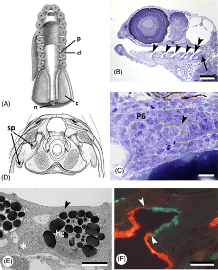
Pouch–cleft contacts and tooth formation. (A) Diagrammatic ventral view of dissected branchial region of an ‘idealised' primitive craniate. c, coelom; cl, cleft; n, notochord; p, pouch [after Bjerring, 1977, with permission from the author and publisher]. (B) Danio rerio; sagittal section showing the open pouches (gill slits) in a 96 h post-fertilisation (hpf) specimen (arrowheads); pharyngeal teeth (arrow) are associated with the last pouch (pouch 6, double arrowhead). Scale bar = 100 μm. (C) Danio rerio; cross section at the level of the forming pharyngeal dentition in a 50 hpf specimen. Note connection of the tooth-forming epithelium (arrowhead) with that lining pouch 6 (P6). Dotted line follows basal lamina, delimiting the pharyngeal epithelium. Scale bar = 10 μm. (D) Latimeria chalumnae; teeth formed in the spiracular pouch (sp); based on a photograph of a transverse section at the level of the intracranial articulation (modified after Millot & Anthony, 1958<, with permission from the publisher). (E) Oryzias latipes; transmission electron microscopy (TEM) picture of a cross section of the pharyngeal epithelium in a 7 days post-fertilisation (dpf) embryo, showing that a flattened cell layer (arrowhead) covers the hatching gland cells (hg) and basal endodermal layer (asterisk) of the pharyngeal epithelium. Scale bar = 5 μm. (F) Danio rerio; sagittal section through the mouth entrance of a 55 hpf Tg(krt4:tomatoCAAX;sox17:egfp) embryo, periderm (red) has partially invaded the mouth (boundary indicated by arrowheads); endoderm is green. Scale bar = 50 μm. |
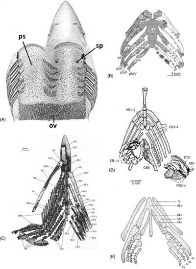
Distribution of pharyngeal teeth. (A) Squalus acanthias; adult pharynx. ov, oesophageal villi; ps, ‘placoid scales’; sp, spiracle (after Cook & Neal, 1921). (B) Polypterus senegalus; visceral skeleton and dentition of pterolarva. bb, basibranchial; cb, ceratobranchial; ch, ceratohyal; eb, epibranchial; hb, hypobranchial; hh, hypohyal; pb, pharyngobranchial (from Wacker et al., 2001, with permission from the publisher). (C) Elops lacerta; visceral skeleton and dentition. Tooth plates associated with (amongst others): BB, basibranchials; HB, hypobranchials; CB, ceratobranchials; EB, epibranchials; PB, pharyngobranchials (after Taverne, 1974). (D) Haemulon sciurus; visceral skeleton and dentition. BH, branchiohyoideus; CB, ceratobranchial; EB, epibranchial; ET, epibranchial tooth plate; HB, hypobranchial; PB, pharyngobranchial [after Wainwright, 2006, with permission from the author and publisher]. (E) Triton alpestris; visceral skeleton and dentition of stage 50, ‘Reusenzähnchen’, structures considered by the author to be true tooth germs. BB, basibranchial; HB, hypobranchial; Hy, hyoid; K, ceratobranchial [from Wagner, 1955; reproduced with permission from the Journal of Embryology and Experimental Morphology]. |
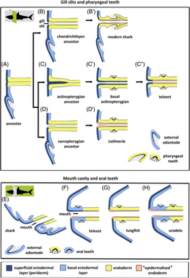
Germ layer contribution to pharyngeal and oral teeth. (A–D′) Hypothetical evolutionary scenario for the persisting influence of ectoderm on pharyngeal tooth formation. (A) Primitive situation; external odontode enamel organ derives from basal ectodermal layer; endodermal pouches contact the ectoderm and endoderm partially spreads under the periderm. (B, B′) In the chondrichthyan lineage, endoderm of the pouches is stratified and the superficial layer is ‘epidermalised'; pharyngeal teeth derive from the basal endodermal layer (modern sharks, B′). (C–C″) In the actinopterygian lineage, periderm invades the pouches, ultimately covering the entire pharyngeal endoderm (basal actinopterygians, C′); in advanced actinopterygians, periderm invasion is restricted to the distal parts of the pouches, while endoderm is ‘epidermalised' medially; pharyngeal teeth derive from the basal endodermal layer (teleosts, C″). (D, D′) In the sarcopterygian lineage, endoderm of the pouches is stratified and the superficial layer is ‘epidermalised'; pharyngeal teeth derive from the basal endodermal layer (Latimeria chalumnae, D′). It is possible that basal actinopterygians took path D, leading to the situation in teleosts (i.e. periderm invasion would be a teleost novelty rather than a regressive feature). A study of basal actinopterygians will resolve this issue. (E–H) Schematic representation of germ layer contribution to oral tooth formation in extant vertebrates, as inferred from literature data. (E) Shark; dental lamina derived purely from ectoderm. (F) Teleost; both periderm and periderm-like layer may constitute the superficial layer of the oral epithelium; enamel organs putatively derived from ectoderm. (G) Lungfish; the oral cavity is lined by endoderm, which also gives rise to the enamel organs. However, the superficial cells have acquired characters similar to cells of the outer epidermal layer. (H) Urodele amphibian; the basal ectodermal layer invades the mouth, oral teeth derive from the basal layer (either ectoderm or endoderm) but are covered by ‘epidermalised' endoderm. Shark profiles show the plane of sectioning (in green): cross sections for gill slits and pharyngeal teeth (upper panels), sagittal sections for mouth cavity and oral teeth (lower panels). |
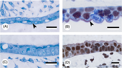
'Epidermalised' endoderm. Detail of the ‘epidermalised' endodermal epithelium (arrowheads) in the roof of the pharyngeal cavity (A, B) and of the ectodermal epithelium of the skin (C, D) in Acipenser ruthenus (A, C) and Ambystoma mexicanum (B, D). Note similarities in differentiation of the superficial cells. Scale bars = 25 μm. |