
|
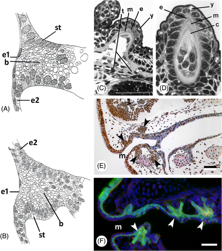
Fig. 6 Germ layer contribution to oropharyngeal epithelium and teeth in lungfish and amphibians. (A) Lepidosiren paradoxa; drawing of a sagittal section of a stage 30 specimen (after Kerr, 1903); drawing mirrored to show anterior to the left. b, yolk-laden cells in the buccal cavity; e1, superficial ectodermal layer; e2, basal ectodermal layer; st, lining epithelium of buccal cavity arising in situ. (B) Ambystoma mexicanum; drawing of a similar section to A, embryo of 7.5 mm length (after Kerr, 1903; drawing mirrored to show anterior to the left). (C, D) Neoceratodus forsteri, cusp of pterygopalatine tooth plate [from Kemp, 2002a, with permission from the author and publisher]. c, circumdenteonal dentine; e, enamel; m, mantle dentine; t, trabeculae; y, yolk remnants. Scale bars: C = 100 μm; D = 20 μm. (E) Ambystoma mexicanum; sagittal section of a stage 43 embryo; compare with F. Tooth buds indicated by arrowheads. m, mouth opening. Scale bar = 50 μm. (F) Ambystoma mexicanum; transgenic embryo of stage 43 with green fluorescent protein (GFP) labelling in the ectoderm [modified from Soukup et al., 2008, courtesy of the authors]. Arrowheads: tooth buds arising in ectoderm; m, mouth opening. Scale bar = 50 μm.