
Fig. 10
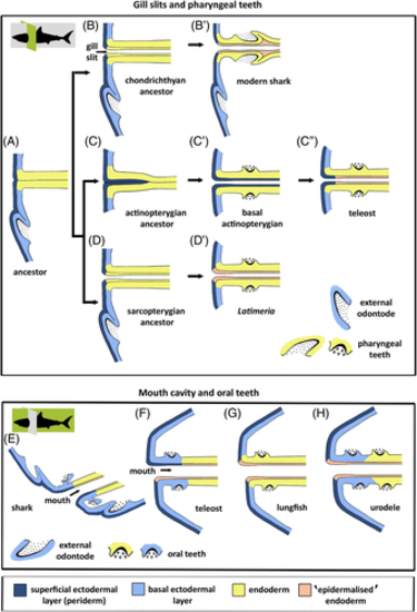
Germ layer contribution to pharyngeal and oral teeth. (A–D′) Hypothetical evolutionary scenario for the persisting influence of ectoderm on pharyngeal tooth formation. (A) Primitive situation; external odontode enamel organ derives from basal ectodermal layer; endodermal pouches contact the ectoderm and endoderm partially spreads under the periderm. (B, B′) In the chondrichthyan lineage, endoderm of the pouches is stratified and the superficial layer is ‘epidermalised'; pharyngeal teeth derive from the basal endodermal layer (modern sharks, B′). (C–C″) In the actinopterygian lineage, periderm invades the pouches, ultimately covering the entire pharyngeal endoderm (basal actinopterygians, C′); in advanced actinopterygians, periderm invasion is restricted to the distal parts of the pouches, while endoderm is ‘epidermalised' medially; pharyngeal teeth derive from the basal endodermal layer (teleosts, C″). (D, D′) In the sarcopterygian lineage, endoderm of the pouches is stratified and the superficial layer is ‘epidermalised'; pharyngeal teeth derive from the basal endodermal layer (Latimeria chalumnae, D′). It is possible that basal actinopterygians took path D, leading to the situation in teleosts (i.e. periderm invasion would be a teleost novelty rather than a regressive feature). A study of basal actinopterygians will resolve this issue. (E–H) Schematic representation of germ layer contribution to oral tooth formation in extant vertebrates, as inferred from literature data. (E) Shark; dental lamina derived purely from ectoderm. (F) Teleost; both periderm and periderm-like layer may constitute the superficial layer of the oral epithelium; enamel organs putatively derived from ectoderm. (G) Lungfish; the oral cavity is lined by endoderm, which also gives rise to the enamel organs. However, the superficial cells have acquired characters similar to cells of the outer epidermal layer. (H) Urodele amphibian; the basal ectodermal layer invades the mouth, oral teeth derive from the basal layer (either ectoderm or endoderm) but are covered by ‘epidermalised' endoderm. Shark profiles show the plane of sectioning (in green): cross sections for gill slits and pharyngeal teeth (upper panels), sagittal sections for mouth cavity and oral teeth (lower panels). |