
Fig. 3
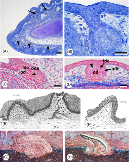
Oropharyngeal epithelium, teeth, and dermal denticles in chondrichthyans. (A) Scyliorhinus canicula; sagittal section of the lower jaw of a 85 mm embryo, showing skin (= dermal) denticles (arrowheads), and the dental lamina with developing teeth (arrows). Scale bar = 100 μm. (B) Scyliorhinus canicula; larger magnification of the early dental lamina with a developing tooth in a 85 mm embryo. Scale bar = 20 μm. (C, D) Scyliorhinus canicula; sagittal section of a tooth anlage in the lower jaw with the invading dental lamina (C) compared to the anlage of a dermal denticle (D) in the same 50 mm embryo. Outer (ode) and inner dental epithelium (ide), the basal lamina of the enamel organ (arrowhead) and the mesenchymal condensation that will give rise to the odontoblasts (od) characterise the anlagen of teeth and dermal denticles. Scale bars = 25 μm. (E, F) Squalus acanthias; two stages of development of a ‘placoid scale’ (from Cook & Neal, 1921). n'v. = nerve; pl. sc. = placoid scale. (G, H) Mustelus sp.; two stages of development of a ‘mucous membrane denticle’ (from Peyer, 1968). |