
Fig. 1
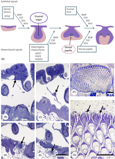
Epithelial primacy in tooth formation. (A) Schematic representation of tooth formation in mammals and transcription factors and signalling molecules involved (modified from Koch, Thesleff & Kreiborg, 2017); purple: epithelium/enamel organ; pink: mesenchyme/dental papilla. BMP, bone morphogenetic protein; FGF, fibroblast growth factor; Shh, sonic hedgehog. (B–E) Intramedullary tooth formation in the lower pharyngeal jaw of the cichlid Hemichromis bimaculatus, 28 mm standard length (SL). Successive cross sections through the epithelial strand (arrow) giving rise to the enamel organ of the replacement tooth, showing its origin (B), its course across the large vascular cavity (C), its penetration through the jaw bone (D) and its distal end within the medullary cavity of the jaw bone (E). Only few mesenchymal cells have condensed around the distal end. Total length of the epithelial strand is approximately 300 μm. Scale bar for B–E = 100 μm. (F, G) Section through the left premaxilla of a scraping loricariid catfish, Ancistrus cf. triradiatus, of 33.5 mm SL. Note virtual absence of mesenchymal cells at the distal end of each successional lamina (arrows). Scale bars: F = 100 μm; G = 50 μm. |