
FIGURE
Fig. 11
Fig. 11
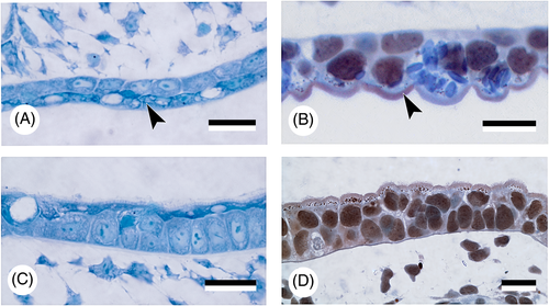
'Epidermalised' endoderm. Detail of the ‘epidermalised' endodermal epithelium (arrowheads) in the roof of the pharyngeal cavity (A, B) and of the ectodermal epithelium of the skin (C, D) in Acipenser ruthenus (A, C) and Ambystoma mexicanum (B, D). Note similarities in differentiation of the superficial cells. Scale bars = 25 μm. |
Expression Data
Expression Detail
Antibody Labeling
Phenotype Data
Phenotype Detail
Acknowledgments
This image is the copyrighted work of the attributed author or publisher, and
ZFIN has permission only to display this image to its users.
Additional permissions should be obtained from the applicable author or publisher of the image.
Full text @ Biol. Rev. Camb. Philos. Soc.