
Fig. 9
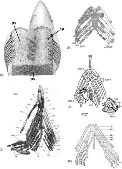
Distribution of pharyngeal teeth. (A) Squalus acanthias; adult pharynx. ov, oesophageal villi; ps, ‘placoid scales’; sp, spiracle (after Cook & Neal, 1921). (B) Polypterus senegalus; visceral skeleton and dentition of pterolarva. bb, basibranchial; cb, ceratobranchial; ch, ceratohyal; eb, epibranchial; hb, hypobranchial; hh, hypohyal; pb, pharyngobranchial (from Wacker et al., 2001, with permission from the publisher). (C) Elops lacerta; visceral skeleton and dentition. Tooth plates associated with (amongst others): BB, basibranchials; HB, hypobranchials; CB, ceratobranchials; EB, epibranchials; PB, pharyngobranchials (after Taverne, 1974). (D) Haemulon sciurus; visceral skeleton and dentition. BH, branchiohyoideus; CB, ceratobranchial; EB, epibranchial; ET, epibranchial tooth plate; HB, hypobranchial; PB, pharyngobranchial [after Wainwright, 2006, with permission from the author and publisher]. (E) Triton alpestris; visceral skeleton and dentition of stage 50, ‘Reusenzähnchen’, structures considered by the author to be true tooth germs. BB, basibranchial; HB, hypobranchial; Hy, hyoid; K, ceratobranchial [from Wagner, 1955; reproduced with permission from the Journal of Embryology and Experimental Morphology]. |