
|
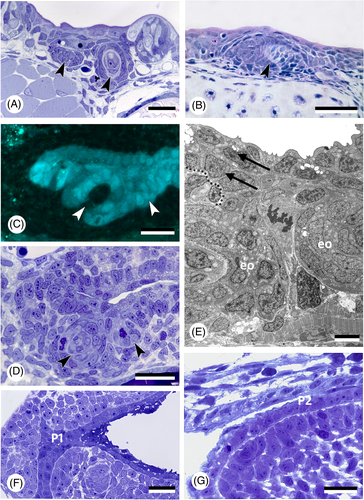
Fig. 4 Oropharyngeal epithelium and teeth in actinopterygians. (A) Polypterus senegalus; pharyngeal epithelium and forming tooth germs (arrowheads) in an 18 mm larva. Scale bar = 20 μm. (B) Acipenser ruthenus; tooth germ (arrowhead) in the buccopharyngeal epithelium in a 15 mm larva. Scale bar = 50 μm. (C, D) Danio rerio, cross section of a 56 h post-fertilisation (hpf) embryo at the level of the first forming teeth (arrowheads), shown in a transgenic (sox17:egfp) line (C), and corresponding semithin, toluidine blue-stained section of a wildtype fish (D). Scale bars = 20 μm. (E) Danio rerio, transmission electron microscopy (TEM) image of the bilayered pharyngeal epithelium (two arrows, delimited by dotted line) of a 72 hpf embryo at the level of the first two teeth (eo, enamel organ). Scale bar = 5 μm. (F) Hemichromis bimaculatus, cross section of the oropharynx of a 1 day post-hatching (dph) (4.0 mm total length) specimen at the level of pouch 1 (P1). Scale bar = 20 μm. (G) Oncorhynchus tshawytscha, sagittal section of the pharyngeal epithelium in an unhatched embryo 17 days post-fertilisation (dpf), at the level of the (open) pouch 2 (P2). Scale bar = 20 μm.