
- Title
-
Aplnra/b Sequentially Regulate Organ Left-Right Patterning via Distinct Mechanisms
- Authors
- Zhu, C., Guo, Z., Zhang, Y., Liu, M., Chen, B., Cao, K., Wu, Y., Yang, M., Yin, W., Zhao, H., Tai, H., Ou, Y., Yu, X., Liu, C., Li, S., Su, B., Feng, Y., Huang, S.
- Source
- Full text @ Int. J. Biol. Sci.
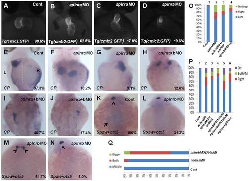
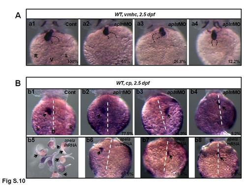
Organ left-right lateral defect in embryos treated with different ways. (A-D, O) The pattern of heart displayed in controls and aplnra/b morphants. Compared with controls (A, 98.6%, n=72), embryos injected with aplnra MO displayed normal-loop (B, 62.5%, n=112; p<0.01), reversed-loop (C, 17.8%, n=112; p<0.01) and no loop (D, 19.6%, n=112; p<0.01). (O, column 1 to 4) The cartogram of heart LR defect for embryos treated with different MOs or mRNA. Only more than 18.2% of aplnrb morphants displayed heart LR defect (O, column 4, n=115; p<0.05). Aplnra mRNA injection (O, column 3, 26.8%, n=71; p<0.05) partially rescued the heart LR defect in aplnra morphants (O, column 2, 37.5%, n=112). (E-J, P). While 97.3% of control embryos showed left-sided expression of cp (E, n=112), 18.2.0% (F, n=176, p<0.01), 9.1% (G, n=176, p<0.05) and 10.8% (P, n=176, p<0.05) of aplnra morphants showed both sided and right-sided expression of cp, or disappear. (H-J, P). Compared with that in aplnra morphants, aplnra mRNA partially rescued liver LR defect (24%, n=121, p<0.05). In aplnra/b double morphants, the expression of cp was greatly downregulated (100%, n=195, p<0.001), and more embryos displayed liver LR defect (H-J, P, 51.3%, n=195, p<0.05) than that in aplnra morphants (P, 38.1%, n=176). In embryos injected with aplnra sgRNAs and Cas9 protein, 15.8%; and 6.4% of them displayed right sided and both sided/middle liver (P, n=233, p<0.05). (K-N, Q). otx5 was expressed in the middle telencephalon. All the embryos injected with Cont MO displayed the expression of otx5 in the middle telencephalon (K, 100%, n=32, and Q, column 1).While 61.7% of embryos injected with aplnrb MO showed both-sided otx5 in telencephalon (M, Q, n=34), only 5.5% of aplnra morphants displayed both-sided otx5 (Q, 37). For observation, heart, ventral view; cp, spaw and otx5, dorsal view. EXPRESSION / LABELING:
PHENOTYPE:
|
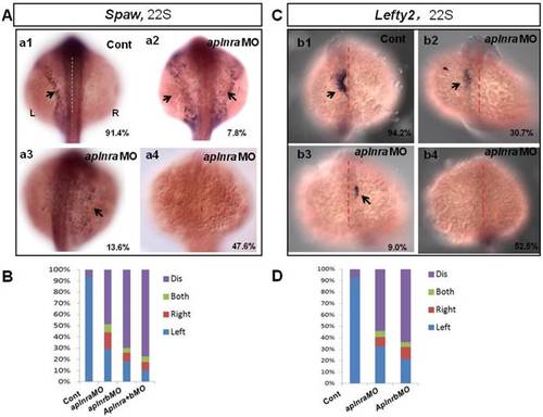
Expression of left-sided Nodal signaling in LPM. (A. a1-a4, B) Expression of left-sided spaw in embryos. In control embryos, near all the embryos expressed left-sided spaw (A. a1, 91.4%, n=93). While in aplnra and aplnrbmorphants, left-sided spaw expression pattern was changed (A. a2-a4, B), 13.6% (p>0.05), 7.8% and 47.6% (p<0.001) of aplnra morphants expressed right-sided, both-sided spaw or spaw disappeared (B, middle-left column, n=103). 10.2 % (p<0.05) and 6.1% (p>0.05) of aplnrb morphants showed right-sided and both sided spaw (B,middle-right column, n=98), majority of aplnrb morphants showed no staining for spaw (B, middle-right column, n=98, p<0.001). (C. c1-c4, D) The expression of left-sided lft2 in the heart progenitor field. In the control embryos, lft2 was expressed in the left side of heart progenitor field (C.c1, 94.2%, n=52). Lft2 was down-regulated in aplnramorphants (C. c2-c4), the left-sided expression pattern was also perturbed (C. c2-c4, D), with 9.0%, 6.3% and 52.5% of embryos showing right-sided, both-sided lft2 and lft2 disappeared, respectively (C. c2-c4; D, middle column, n=74). The lft2 expression pattern in aplnrb morphants (D, right column, n=93) was similar to that in aplnramorphants (D, middle column, n=74), but more embryos had no staining for lft2 in aplnrb morphants (D, right column, n=93) than in aplnra morphants (D, middle column, n=74). All the embryos were observed from dorsal view. L, Left; R, Right. EXPRESSION / LABELING:
PHENOTYPE:
|

ZFIN is incorporating published figure images and captions as part of an ongoing project. Figures from some publications have not yet been curated, or are not available for display because of copyright restrictions. PHENOTYPE:
|
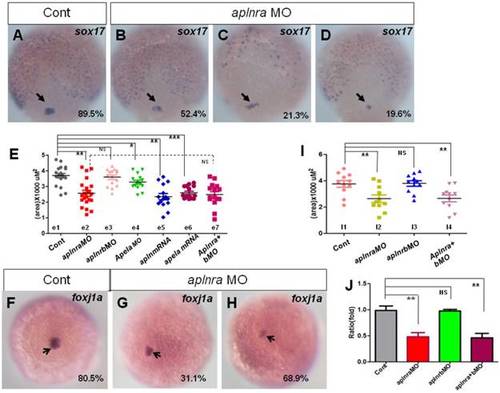
Expression of sox17 and foxj1a. (A-E) Expression of sox17 in DFCs at 90% epiboly. In control embryos, 89.5% of embryos showed normal expression (A, n=88). In aplnra morphants, three kinds of expression pattern were discovered: mild decreased (B, 52.4%, n=89, p<0.001), scattered (C, 21.3%, n=89, p<0.001) and decreased (D, 19.6%, n=89, p<0.01). The area of sox17 expression was measured and in control embryos, the average level was 3.68x10³ uM2 (E. e1 group, n=19), the average area in aplnra morphants was 2.55x10³ uM2 (E. e2 group, n=22, p<0.05). Sox17 expression areas were also measured for aplnrb morphants, aplnra+b morphants, apela morphants, embryos injected with apln mRNA and embryos injected with apln mRNA, respectively (E, e3-e7 group). The average areas were 3.63x10³ uM2 (E, e3 group, aplnrb morphants, n=15, p>0.05), 2.53x10³ uM2 (E. e7 group, aplnra+bmorphants, n=15), 3.28x10³ uM2 (E, e4 group, apela morphants, n=15, p<0.05), 2.46x10³ uM2 (E, e5 group, apln mRNA, n=15, p<0.01) and 2.64x10³ uM2 (E, e6 group, apela mRNA, n=15, p<0.001), these data indicated sox17expression was down reglated in all these treated embryos, excepted for aplnrb morphants. (F-J) Analysis of foxj1aexpression. Foxj1a was expressed in DFCs at 90% epiboly in control (F) and treated embryos (G, H). Compared with control embryos (F, 80.5%, n=82), foxj1a was greatly down regulated in aplnra morphants (H-I, 68.9%, n=87, p<0.001) as well in aplnra+b morphants (I, 65.7%, n=76, p<0.001), but not in aplnrb morphants (I, 18.9%, n=74, p>0.05). Foxj1a expression area in control embryos, aplnra morphants, aplnrb morphants and aplnra+b morphants were average 3.75 x10³ uM2. 2.56 x10³ uM2, 3.57 x10³ uM2 and 2.39 x10³ uM2 respectively (I). Compared with that in control embryos, q-PCR experiment showed the quantity of foxj1a expression in aplnra morphants and aplnra+bmorphants were down-regulated with 0.48 folds and 0.46 folds respectively, while the expression of foxj1a in aplnrbmorphants was not affected (J). *, p<0.05; **, p<0.01; ***, p<0.001; NS, not significant. EXPRESSION / LABELING:
PHENOTYPE:
|
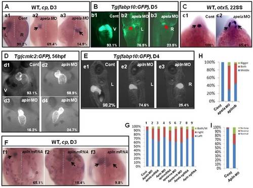
Organ LR asymmetry defect in embryos with aplnr ligands loss or gain of function. (A, G) Majority of control embryos expressed left-sided liver marker cp in day 3 (A. a1, G. column 1, 90.2%, n=51). Liver LR asymmetry was disturbed in apela MO injected embryos (A. a2-a3, G. column 2, 30.6%, n=121, p<0.01) and apela mRNA injected embryos (G. column 3, 34.9%, n=103, p<0.01). In transgenic line Tg(fabp10:GFP), the liver LR defect was also observed, displayed left-sided and right-sided liver in 76.5% and 23.5% of apela morphants, respectively (B. b2 and b3, n=51), but the GFP in liver region started to express in day 5 (B. b2 and b3). (C) Head marker otx5 was expressed in the middle telencephalon (C. c1, H. column 1, 100%, n=32), but many of apela morphants (C. c2, H. column 2, 65.4%, n=26) and aplnrb morphants (H. column 3 61.7%, n=34) showed both-sided otx5 in telencephalon. (D, I) Apln loss of function resulted heart LR asymmetry defect (D. d2-d4, I), displayed reversed loop (D. d4, I, 24.7%, n=117, p<0.05), normal loop (D. d2, I, 58.9%, n=117, p<0.01) and linear heart (D. d3, I, 16.2%, n=117, p<0.05). (E. e1-e3, G) In apln morphants, liver development was not delayed (E), but 25.4% liver LR asymmetry defect was found (E. e2-e3, G. column 4, n=185, p<0.001). Apela mRNA or apln mRNA gain of function also resulted in liver LR asymmetry defect, 34.9% (p<0.01) and 37.2% (p<0.01) of embryos displayed right-sided or both-sided expression of cp (G. column 5, n=97). When compared with the liver LR defect in apela morphants (G. column 2, 30.6%, n=121), high ratio of embryos co-injected with apela MO and aplnra MO (G. column 6, 37.1%, n=70), or embryos co-injected with apela MO and aplnrb MO (G. column 7, 40.0%, n=91) showed liver LR asymmetry defect. In embryos injected with apela/apln sgRNAs with Cas9 protein, the livers also showed left right asymmetry defect (G. column 8 and 9). L, left; R, right; V, ventral view; D3, day 3; D5, day 5. EXPRESSION / LABELING:
PHENOTYPE:
|
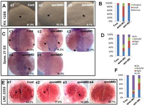
KV morphogenesis and left-sided Nodal signaling in apela morphants. (A-B) In control, 91.2% of embryos showed normal KV (A, n=136), but 37.6%, 53.3% and 9.1% of embryos injected with apela MO showed normal KV, mild smaller KV and no KV, respectively (B, C and D. column2, n=178). (C. c1-c5, D) Majority of control morphants expressed left-sided spaw in the LPM (C. c1, D left column, 91.4%, n=35). In apela MO morphants, 31.3%, 13.9% and 16.3% of embryos showed left-sided spaw, right-sided spaw and both-sided spaw (C. c2-c4, D middle column, n=30), while the spaw expression in most of apela morphants couldn't be found (C. c5, D middle column, 38.9%, n=86). (E-F) The Nodal downstream gene lft2 was checked. Compared with that in control embryos (E. e1, F right column, 94.2%, n=52), lft2 was down regulated and the left-sided expression pattern was disturbed in apelamorphants (E. e2-e4, F middle column, n=39), displaying left-sided (17.9%), right-sided (10.25%), both-sided (5.12%) expression and the expression disappeared (66.7%). The disturbed KV morphogenesis and the perturbed spaw or lft2 expression were also observed in apln morphants (D, D, F, right column). EXPRESSION / LABELING:
PHENOTYPE:
|
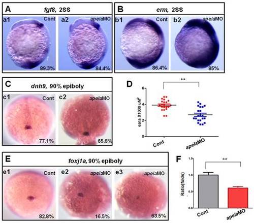
KV/cilia related signaling pathway analysis in apela morphants. (A-C) Compared with the control embryos (A. a1, B. b1 and C. c1), the expression of fgf8 and the downstream gene dnh9 were not changed in apela morphants (A. a2, 84.4%, n=32; C. c2, 65.6%, n=32), but the expression of erm was up regulated in apela morphants (B. b2, 85%, n=20). (E-D) Foxj1a was expressed in DFCs. Compared with that in control embryos (E. e1, 82.8%, n=35), foxj1awas down regulated in apela morphants (E. e3, 63.5%, n=104). The average area of foxj1a expression in control embryos and apela morphants were 3.87 x10³ uM2 (n=21) and 2.75 x10³ uM2, respectively (D, n=21, p<0.01). Q-PCR experiment indicated that foxj1a expression was down regulated with 0.61 folds compared with that in control embryos (F). *, p<0.05; **, p<0.01. EXPRESSION / LABELING:
PHENOTYPE:
|
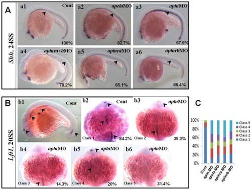
Expression of midline related genes in different treated embryos. (A. a1-a6) Shh expression at 24SS. Compared with that in control embryos (A. a1, 100%, n=31), no clearly decreased or increased expression of shh in midline was found in apela morphants (A. a2, 82.7%, n=29), aplnra+b double morphants (A. a4, 78.2%, n=23), aplnra morphants (A. a5, 85.1%, n=27) and aplnrb morphants (A. a6, 86.4%, n=22). In apln morphants, the expression of shh was slightly increased (A. a3, 67.9%, n=28). (B. b1-b6, C) Expression of lft1 at 20SS. In wild type embryos lft1 was expressed in 4 domains, including left telencephalon, left heart field, trunk midline and tail midline (B. b1, black arrow head, n=45), and in dorsal view, lft1 was found to be expressed in left telencephalon, left heart field, trunk midline (B. b2, black arrow head). In apln morphants, lft1 expression was found to be decreased with different phenotypes (B. b3-b6, n=85). Among all the alpn morphants, lft1 in midline was disappeared or decreased greatly in more than half of embryos (B. b4-b6, class 3 to class 5; C, the second column, 64.7%, n=85, p<0.01). The expression pattern of lft1 in apln morphants was also found in apela morphants (C, column 3, 58.2%, n=79, p<0.01), aplnra morphants (C, column 4, 60%, n=75, p<0.01) and aplnrb morphants (C, column 5, 56.2%, n=89, p<0.01), near or more than half of embryos showed greatly decreased lft1 in the trunk midline. EXPRESSION / LABELING:
PHENOTYPE:
|
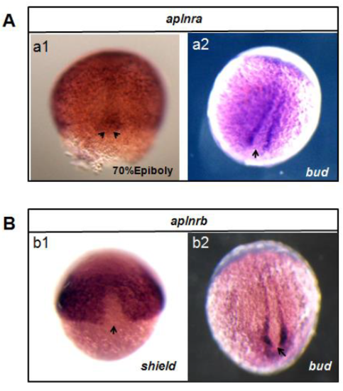
The expression pattern of aplnra/aplnrb at gastrulation stage and bud stage (A) The expression pattern of aplnra. aplnra is expressed ubiqiuously at 70% epiboly, it is also expressed in DFCs(a1, arrow head). At bud stage, it is expressed in the cells near midline while not in KV progenitors(a2, arrow). (B) The expression pattern of aplnrb. aplnrb is not expressed in the DFCs at shield stage (b1, arrow), the expression pattern is similar to that of aplnra at bud stage (b2). |
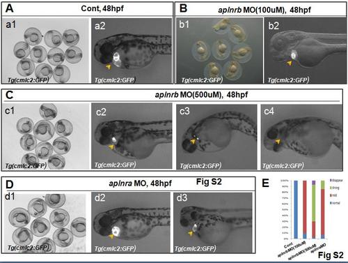
: The heart phenotype for aplnrbor aplnraloss of function (A-E) The heart development in controland aplnra/bmorphants. Compared with the controlmorphants (A. a2, E, 100%, n=57), the heart in most of embryos injected with aplnrbMO(100uM) displayed mild smaller (B. b2, E, 89.3%, n=103), whilein most of aplnrbmorphants(500uM), the heart became very small (C. c3, E, 63.6%, n=99) orthe heart disappeared (C. c4, E,27.3%, n=99), only small part of embryos had the mild smaller heart (C. c2, E, 7.1%, n=99). For aplnra morphants, the phenotype was not strong, majority of embryos displayed mild smaller heart (D.d2, E, 78.7%, n=108), only smaller number of embryos displayed very small heart (D. d3, E, 14.8%, n=108). PHENOTYPE:
|
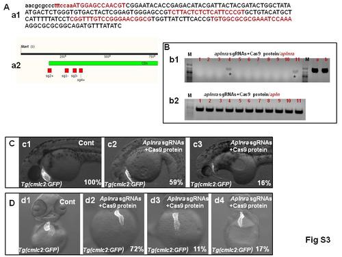
The heart phenotype of embryos injected with aplnra sgRNAs and Cas9 protein (A. a1-a2) The four target points for aplnra gene. a1 showed the detailed sequence for each targeting point in the genomic DNA. a2 showed the location of the target points in the genome map. (B. b1-b2) The efficiency of genome editing using CRISPR/Cas9 system. The sorted embryos injected with aplnra sgRNAs and Cas9 protein were used to prepare the genomic DNA, PCR experiment showed that the genomic DNA in control embryos was intact (B. b1, line a and b), while the genomic DNA was edited successfully in nearly all the embryos (88.9%, n=54), 11.1% of embryos' genomic DNA was not completely edited (cutting off part of target genome) (B. b1, line 1-11, stars showed, n=27, amplified with aplnra primers). Using the genomic DNA from the embryos injected with sgRNAs and Cas9 protein as templates, the apln can be amplified (B. b2, line1-11), meaning all the templates worked well. (C) The heart progenitor number decreased in most of embryos injected with aplnra sgRNAs and Cas9 protein (c2 showed the embryos with mild decreased heart number, c3 showed the embryos with heart number decreased greatly.) (D) The heart left right asymmetry defect in embryos with mild heart number decreased (D. d2-d4, n=89). |
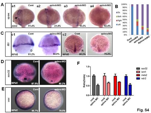
The expression of nodal related genes in aplnra morphants (A-B) The expression of spaw in control embryos and aplnra morphants. In control, 91.4% of embryos displayed left-sided spaw(A. a1 and B, n=93), 21.4%, 10.2% and 62.4% of embryos injected with aplnrb MO(500uM) displayed left-sided spaw, right-sided spaw and spaw disappeared (A. a2-a4 and B, n=98). (C) Compared with control embryos, at 60% epiboly, 84.8% of embryos injected with aplnra MO displayed mild decreased lft1 expression (Cc1, n=33), at 95% epiboly, 79.4% of embryos injected with aplnra MO displayed greatly decreased lft1 expression (Cc2, n=33). (D) The expression of sox17 indicated that the transcription level of sox17 was down-regulated in aplnra morphants (80.2%, n= 26), most of control embryos displayed high level of sox17 (87.5%, n=24). (E) In control, 95.7% of embryos displayed high level of vox expression (n=23), while vox was down-regulated in 78.5% of embryos injected with aplnra MO (n=28). (F) Q-PCR analysis for the nodal related genes sox32, ism1, ndnr2 and mxtx2 in control and aplnra morphants. The result indicated that all the four genes were down-regulated in aplnra morphants. Note: L, Left; R, Right; D, Dorsal; V, Ventral; Three repeated experiments were done for Q-PCR. *, P<0.05; **, P<0.01; NS, not significant difference. Fig. |
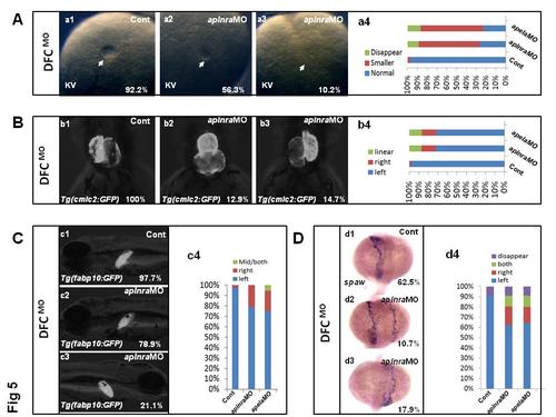
aplnra or apela loss of function in DFCs resulted in LR patterning defect. (A. a1-a4) The KV phenotype in control and aplnra or apela loss of function in DFCs. At 10 somite stage, 56.3% and 10.2% of embryos injected with aplnra MO in DFCs displayed smaller KV and disappeared KV(A. a2-a4, n=119), 57.6% and 12.0% of embryos injected with apela MO in DFCs displayed smaller KV and disappeared KV (A. a4, n=90), while 92.2% of control embryos displayed normal KV (A. a1 and a4, n=103). (B. b1-b4) At 60hpf, part of embryos injected with aplnra MO displayed linear heart (B. b2 and b4, 14.7%, n=116) and reversed heart (B. b3 and b4, 12.9%, n=116), 15.5% and 13.1% of embryos injected with apela MODFCs displayed reversed heart and linear heart (B. b4, n=90). (C. c1-c3) Liver LR defect was also observed in aplnra MO (total 21.1%, n=95) or apela MO (total 24.8. %, n=90) injected embryos in DFCs (C. c3 and c4). (D. d1-d4) spaw expression was examined in embryos, right-sided and both-sided spaw was discovered in 17.9% and 10.7% of embryos with aplnra loss of function in DFCs (D. d2-d4, n=56) or 15.5%, 11.1% and 8.9% of embryos displayed right-sided, both-sided and disappeared spaw with apela loss of function in DFCs(Dd4, n=56) |
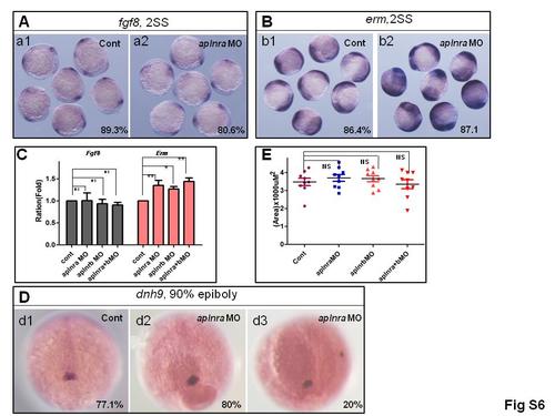
The expression of somecritical regulators being related to LR patterning. (A, B)The expression of fgf8and erm. No difference was observed for fgf8expression in controlembryos and aplnramorphants at 2 somite stage (A. a1-a2, n=36). But erm, the downstream gene of fgf8, was up regulated in aplnramorphants (B. b1-b2, n=31). (C)The expression of fgf8and erminthe control embryos, aplnramorphants, aplnrbmorphants and aplnra+bmorphants were checked by Q-PCR.The result demonstrated that, among them, no difference was observed for fgf8expression, but all the morphants displayed increased expression of erm. (D, E) The expression of dnah9in embryos treated differently. Compared with that in control (D. d1, n=35), the expression of dnh9was not changedgreatlyin aplnramorphants (D. d2, d3, n=30), this result was confirmed by measuring the area in control embryos and aplnramorphants (E). The expression of dnh9was also checked in aplnrband aplnra+bmorphant, no distinct difference was observed among these embryos (E).Note: *, P<0.05; **, P<0.01; NS, not significant difference. |
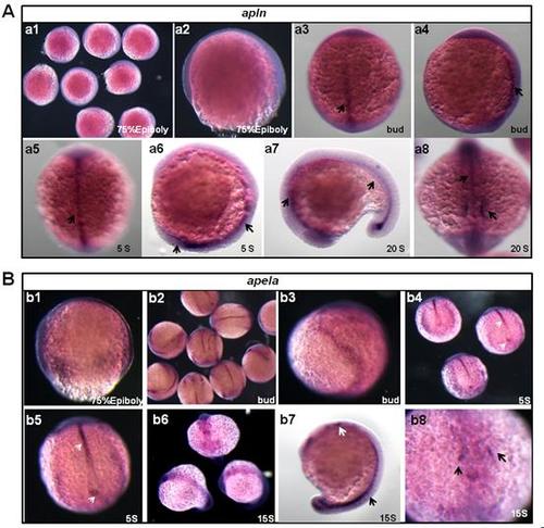
The expression of apln and apela at different stages. (A. a1-a8) The expression of apln from 75% E to 20 SS. At 75% E, apln was not expressed in the embryos (A. a1-a2). At bud to 5 SS, apln was expressed in the midline (A. a3-a6, arrow showed). At 20SS, apln was expressed in midline (A. a7, right arrow; a8, up arrow) and heart progenitor field (A. a7, left arrow; a8, down arrow). (B. b1-b8) The expression of apela from 75% E to 15 SS. At 75% E, apela was expressed ubiquitously (B. b1). At bud stage, apela was expressed in the midline and presomite mesoderm (PSM) (B. b2-b3). At 5SS, apela was expressed in the midline and KV epithelium (B. b4-b5, arrow head). At 15SS, apela was expressed in midline and heart progenitor field (B. b6-b8, arrow). EXPRESSION / LABELING:
|
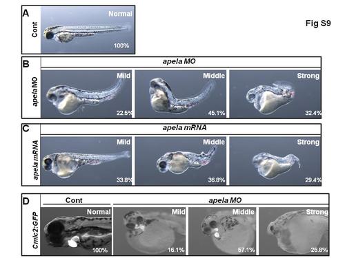
: The similar phenotypes between embryos injected with apelamRNA or apelaMO (A-C) The living embryos injected with controlMO, apelaMO or apelamRNA. At 2 days post fertilization, embryos injected with control MO developed normally (A, 100%, n=45). The embryos injected with apelaMO or apelamRNA had the similar phenotype, displayed mild(22.5%in apelaMO, n=71; 33.8% in apelamRNA, n=68), middle(45.1% in apela MO, n=71; 36.8% in apela mRNA, n=68) and strong phenotype(32.4% in apela MO, n=71; 29.4% in apela mRNA, n=68) (B, C). Meanwhile, the heart cell number was also decreased greatly in apelaMO injected embryos (D, n=56). PHENOTYPE:
|
The organ LR patterning defect in apln morphants and apela mRNA injected embryos. (A. a1-a4) The heart LR patterning in control and apln MO injected embryos. In control embryos, 100% of embryos displayed normal heart loop (n=112). In apln morphants, 61%, 26.8% and 12.2% of embryos displayed normal, reversed and linear heart (A. a2-a4, n=41). For liver LR patterning, 97.3% of control embryos displayed left liver (B. b1, n=112). In apln morphants, 77.5%, 14.3% and 8.2% of embryos displayed left-sided, right-sided and middle/both sided liver (B. b2-b4, n=49). In embryos injected with apela mRNA, most of them displayed abnormal development (B. b5, arrow showed), 61.5%, 18.4% and 9.8% of embryos displayed left-sided, right-sided and middle/both sided liver (B. b6-b8, n=103). |
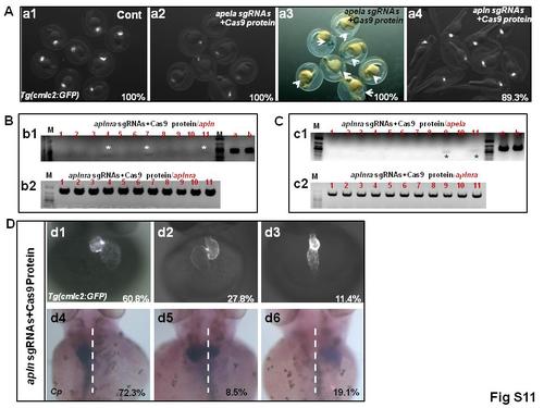
The phenotype in embryos co-injected with apela/apln sgRNAs and Cas9 protein. (A. a1-a4) The heart phenotype in living embryos co-injected with apela/apln sgRNAs and Cas9 protein. In control embryos, 100% of them had normal heart (A. a1, n=107). In embryos co-injected with apela sgRNAs and Cas9 protein, all the embryos displayed very smaller heart or heart disappear (A. a2, 100%, n=136), meanwhile all the embryos displayed intumescent cardiocoelom (A. a3, arrow head, 100%, n=136. Note that a2 and a3 are the same sample). In embryos co-injected with apln sgRNAs and Cas9 protein, no difference about heart size was observed (89.3%, n=141). (B, C) apela and apln genomic DNA was edited by co-injected Cas9 protein and sgRNAs. (D. d1-d6) The heart and liver LR patterning defect in embryos injected with Cas9 protein and apln sgRNAs. There were 60.8%, 27.8% and 11.4% of the embryos showed normal looped heart, reverse looped heart and linear heart (D. d1-d3, n=79), 72.3%, 8.5% and 19.1% of them displayed left-sided, middle and right-sided liver (D. |
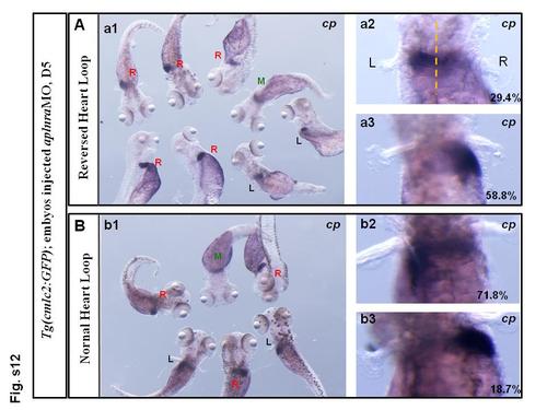
Fig.S12 The heart and liver LR patterning defect in aplnramorphants did not occur at the same time. (A. a1-a3) The liver LR patterning for the embryos displayed reversed loop heart in aplnramorphants. Of them, 29.4% of embryos displayed left-sided liver (A. a2, n=17); 58.8% of the embryos displayed right-sided liver (A. a3, n=17). (B. b1-b3) The liver LR patterning for the embryos displayed normal loop heart in aplnramorphants. Of the embryos, 71.8% of the embryos displayed left-sided liver (B. b2, n=32); 18.7% of embryos displayed right-sided liver (B. b3, n=32). |