
- Title
-
Cystic kidney gene seahorse regulates cilia-mediated processes and Wnt pathways
- Authors
- Kishimoto, N., Cao, Y., Park, A., and Sun, Z.
- Source
- Full text @ Dev. Cell
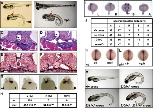
Phenotypes Caused by Defective seahorse (A) A seahorsehi3308 mutant on 5 dpf showing kidney cysts (arrow) and body curvature. (B) A seahorse morphant on 3 dpf showing kidney cysts (arrow) and body curvature. An uninjected control sibling is also shown. (C and D) Cross-section of the glomerular-tubular region of a wild-type and a seahorsehi3308 mutant embryo at 50 hpf. Arrows point to the glomerular and tubular region. (E and F) Increased diameter of the pronephric duct (arrows) in a seahorsehi3308 mutant at 50 hpf, compared to a wild-type embryo. (G–L) seahorse and left-right asymmetry. (G) In situ hybridization for cardiac myosin light chain (cmlc2) on embryos at 26 hpf showing the position of the heart tube. All are ventral views with head to the top. (H) Tabulation of heart position of embryos from three independent crosses. (I) In situ for spaw expression in lateral plate mesoderm on embryos at the 20-somite stage in dorsal views with head to the top. (J) Tabulation of spaw expression pattern. (K and L) The midline structure is not affected by sea-MO as shown by shh expression (K) and ntl expression (L) in embryos fixed at the 8-somite stage. Both in situ are shown in dorsal views with head to the top. (M) seahorsehi3308 and ift172hi2211 double mutants show similar phenotypes as single mutants. All embryos are at 4 dpf, shown in side views with head to the left. Phenotypic embryos (body curvature and kidney cyst) are shown in all crosses except for the wild-type cross. wt, wild-type; mut, mutant; mph, morphant; ctr, embryos injected with the control morpholino oligo; nc, notochord; gl, glomeruli; L, left side; M, middle; R, right side; A, absent; C-MO, embryos injected with a standard control oligo; sea-MO, embryos injected with seahorse morpholino; n, number of embryos analyzed. EXPRESSION / LABELING:
PHENOTYPE:
|
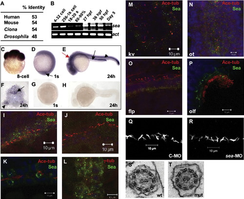
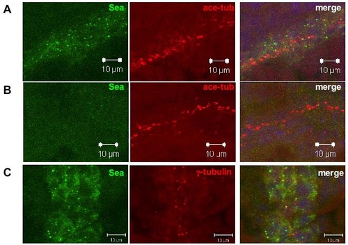
seahorse Is Enriched in Ciliated Tissues, but Is Not Required for Cilia Assembly or Motility (A) Percentage of sequence identity between zebrafish Seahorse and homologs of Seahorse from multiple species. (B) RT-PCR time course for seahorse expression during early development. S, somite; %, percent epiboly; sea, seahorse; act, β-actin. (C–H) In situ hybridization for seahorse on embryos fixed at different stages. (C) Side view of an eight-cell stage embryo with animal pole to the top. (D) Side view of an embryo at one-somite stage with anterior to the top and dorsal to the right. Arrow points to Kupffer's vesicle. (E) Side view of an embryo at 24 hpf with head to the left and dorsal to the top. Red arrow points to the otic vesicle, black arrow points to the pronephric duct. Black arrowhead points to the floor plate. (F) Cross-section of a 24 hpf embryo with dorsal to the top. Arrow points to the floor plate while arrowhead to the pronephric duct. (G) Sense control of (D). (H) Sense control of (E). (I–L) Confocal projections of immunostaining in the pronephric duct. Wild-type (I) and sea morphants (J) are shown at the 18-somite stage; the wild-type embryo in (K) is 50 hpf. Seahorse signal is shown in green, and acetylated tubulin (cilium) in red. (L) Wild-type embryo at 18-somite stage, with Seahorse in green and γ tubulin (centrosome/basal body) in red. (M–P) Confocal projections showing Seahorse in green and acetylated tubulin in red. (M) Kupffer's vesicle (kv) in an 8-somite stage embryo. (N) The otic vesicle (ot) in an embryo at 24 hpf. (O) The floor plate (flp) in an embryo at 24 hpf shown in a side view with anterior to the right and dorsal to the top. (P) The olfactory pit (olf) in an embryo at 78 hpf. (Q and R) Confocal images showing cilia labeled with anti-acetylated tubulin in embryos injected with control or seahorse MO at the 20-somite stage. (S and T) Electron micrographs showing cross-sections of the cilium. C-MO, embryos injected with control morpholino; sea-MO, embryos injected with seahorse morpholino; wt, wild-type; mut, mutant. EXPRESSION / LABELING:
PHENOTYPE:
|
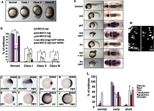
seahorse and Wnt Signaling (A) Wild-type and seahorse morphants at early segmentation stage. All are side views with anterior to the top and dorsal to the right. Class I has mild precocious tail extension. Class II has severe tail extension but with eyecups. Class III has severe tail extension and reduction of eye cups. (B) Embryos injected with indicated combination of morpholino oligos and mRNA from three independent experiments. *p < 0.05. (C and D) Side view of chordin expression at late-shield stage. Dorsal is to the right. (E–J) Animal pole views with dorsal to the right. (E and F) Expression of gsc at 30% epiboly. (G–J) Expression of gsc and vent at shield stage. Ctr, embryos injected with the control morpholino; mph, embryos injected with sea-MO. (K) Embryos injected with 1 ng sea-MO (sea), 0.1 ng prickle 1 (pk1) morpholino and 1 ng inversin (inv) morpholino in different combinations as shown in the figure. Left panel shows embryos on 1 dpf in side views with head to the left. Right panel shows in situ for krox20 and myoD on embryos fixed at the early segmentation stage, all are flat-mounted in dorsal views with head to the left. (L) Tabulation of embryos with body curvature and short body axis phenotypes. The “short” phenotype refers to embryos with only residual tail extension or residual yolk extension. *p < 0.001 for sea + pk1 (n = 3), sea + inv (n = 3), and inv + pk1 (n = 3) when compared with samples injected with sea (n = 4), pk1 (n = 4), or inv (n = 6) morpholino alone. (M) Paraxial cells labeled with membrane-anchored eGFP. Embryos are in dorsal views with anterior to the top and midline to the right. Left panel shows an embryo injected with control morpholino (C), while right panel shows an embryo injected with 2 ng sea-MO. EXPRESSION / LABELING:
PHENOTYPE:
|
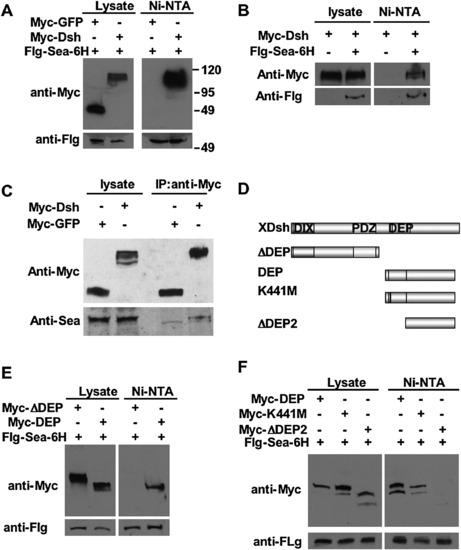
Biochemical Interaction between Seahorse and Dsh Upper: Western blot with anti-Myc on lysates directly or samples pulled down with Ni-NTA (A and B) or anti-Myc (C). Lower: Western analysis with anti-Flag (Flg) (A and B) or anti-Seahorse (C). (A) Flag and 6x His-tagged Seahorse (Flg-Sea-6H) was co-overexpressed with Myc-tagged GFP (Myc-GFP) or Dsh (Myc-Dsh). (B) Myc-tagged Dsh (Myc-Dsh) were overexpressed in the presence or absence of overexpressed 6x His-tagged Seahorse. (C) Myc-tagged GFP (Myc-GFP) or Dsh (Myc-Dsh) was overexpressed in the absence of overexpressed Seahorse. A faster migrating band could also been seen in the lower panel of pull-downs by anti-Myc. Since this band migrated faster than Seahorse and since it was present in both samples, it is likely to be nonspecific. (D) Diagram of Dsh domain structure and different deletion and mutation constructs. (E and F) Different versions of Myc-tagged Dsh were co-overexpressed with Flag and 6x His-tagged Seahorse (Flg-Sea-6H). Upper: Western blot with anti-Myc on lysates directly or samples pulled down with Ni-NTA. Lower: Western analysis with anti-Flag on lysates. |
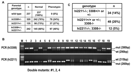
Identification of ift172 seahorse Double Mutants PHENOTYPE:
|
Details for Seahorse Subcellular Localization EXPRESSION / LABELING:
PHENOTYPE:
|
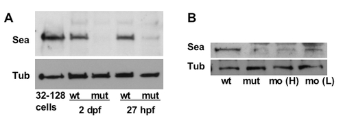
(A) Western blots with anti-Seahorse (upper panel, Sea) and anti-tubulin (lower panel, Tub) on lysates from embryos at 32-128 cell stage, wild type siblings (wt) and seahorsehi3308 mutants (mut) on 2 dpf and 27 hpf. (B) Western blots with anti- Seahorse (upper panel, Sea) and anti-tubulin (lower panel, Tub) on lysates from wild type siblings (wt), seahorsehi3308 mutants (mut), embryos injected with 2 ng (mo (H)) and 1 ng seahorse (mo (L)) morpholino on 1 dpf. |

Unillustrated author statements PHENOTYPE:
|
Reprinted from Developmental Cell, 14(6), Kishimoto, N., Cao, Y., Park, A., and Sun, Z., Cystic kidney gene seahorse regulates cilia-mediated processes and Wnt pathways, 954-961, Copyright (2008) with permission from Elsevier. Full text @ Dev. Cell