
Fig. 3
|
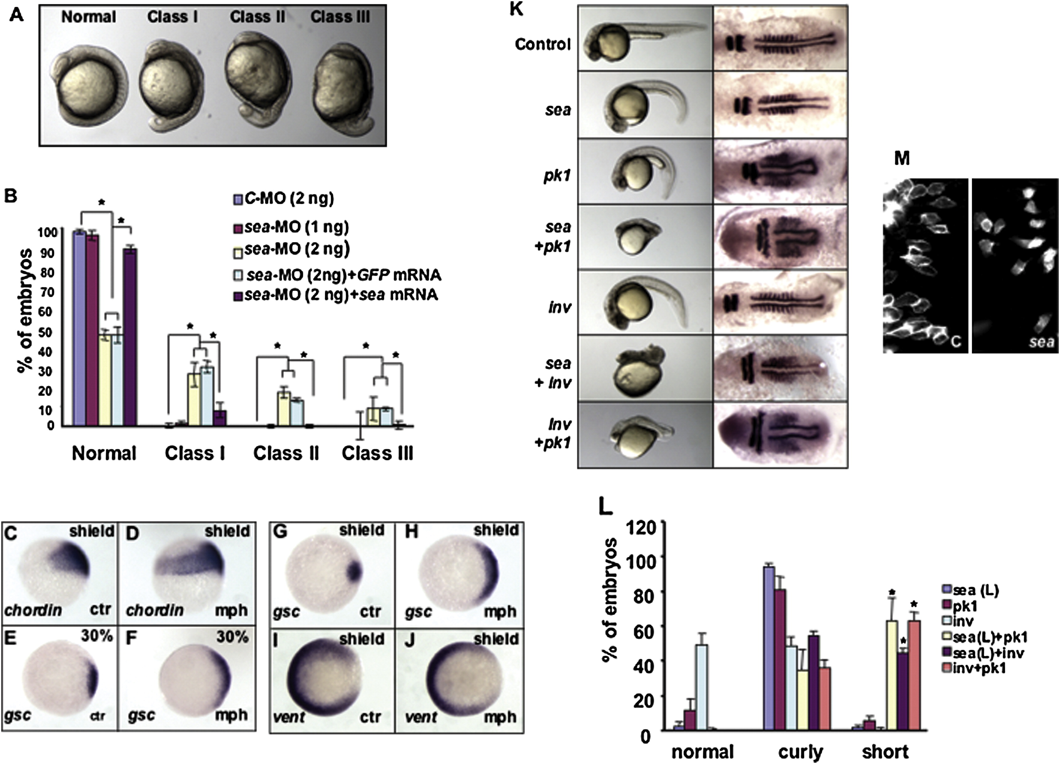
Fig. 3 seahorse and Wnt Signaling
(A) Wild-type and seahorse morphants at early segmentation stage. All are side views with anterior to the top and dorsal to the right. Class I has mild precocious tail extension. Class II has severe tail extension but with eyecups. Class III has severe tail extension and reduction of eye cups.
(B) Embryos injected with indicated combination of morpholino oligos and mRNA from three independent experiments. *p < 0.05.
(C and D) Side view of chordin expression at late-shield stage. Dorsal is to the right.
(E–J) Animal pole views with dorsal to the right. (E and F) Expression of gsc at 30% epiboly. (G–J) Expression of gsc and vent at shield stage. Ctr, embryos injected with the control morpholino; mph, embryos injected with sea-MO.
(K) Embryos injected with 1 ng sea-MO (sea), 0.1 ng prickle 1 (pk1) morpholino and 1 ng inversin (inv) morpholino in different combinations as shown in the figure. Left panel shows embryos on 1 dpf in side views with head to the left. Right panel shows in situ for krox20 and myoD on embryos fixed at the early segmentation stage, all are flat-mounted in dorsal views with head to the left.
(L) Tabulation of embryos with body curvature and short body axis phenotypes. The “short” phenotype refers to embryos with only residual tail extension or residual yolk extension. *p < 0.001 for sea + pk1 (n = 3), sea + inv (n = 3), and inv + pk1 (n = 3) when compared with samples injected with sea (n = 4), pk1 (n = 4), or inv (n = 6) morpholino alone.
(M) Paraxial cells labeled with membrane-anchored eGFP. Embryos are in dorsal views with anterior to the top and midline to the right. Left panel shows an embryo injected with control morpholino (C), while right panel shows an embryo injected with 2 ng sea-MO.
Reprinted from Developmental Cell, 14(6), Kishimoto, N., Cao, Y., Park, A., and Sun, Z., Cystic kidney gene seahorse regulates cilia-mediated processes and Wnt pathways, 954-961, Copyright (2008) with permission from Elsevier. Full text @ Dev. Cell